Methods
Passmore Lab
Methods
Passmore Lab

An integrated approach to understand mechanism
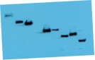
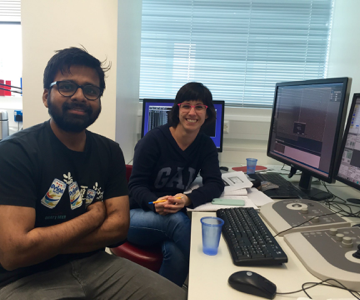
We often start by purifying native protein complexes from the yeast Saccharomyces cerevisiae using TAP tagging approaches, or using CRISPR-Cas9 to tag endogenous genes in human cells. This allows us to determine the architecture of complexes and characterise their functions. We then establish recombinant expression of the protein complexes in insect cells. Once we have high-quality, purified protein, we study it using electron cryo-microscopy (cryoEM), as well as x-ray crystallographic, NMR, biophysical, biochemical and genetic methods. Cross-linking mass spectrometry also plays a major role in understanding the architecture and structure of complexes.
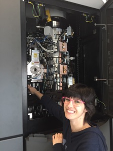
The MRC Laboratory of Molecular Biology electron microscopy facility houses a number of electron microscopes, including four 300 keV Titan Krios, a Glacios, two Polaras, an F20, and microscopes for sample preparation. The Passmore lab developed new gold and graphene specimen supports that improve cryo-EM images.
Importantly, we do not let methods define our work - we use whatever technique we need to in order to understand the biological problems we are addressing. This often involves collaboration with other groups and sometimes requires us to push the boundaries of what is possible.
We ran cryo-EM courses in 2014, 2017 and 2023 as an introduction to the method. The lectures are available online from the following link:
https://www2.mrc-lmb.cam.ac.uk/research/scientific-training/
Passmore Lab, MRC Laboratory of Molecular Biology, Cambridge UK