Amphiphysin and arfaptin BAR domains
This material is supplemental to Peter et al., "BAR Domains as Sensors of Membrane Curvature: The Amphiphysin BAR Structure" Science Express Nov 26 (abstract); print version Jan 23, 2004.
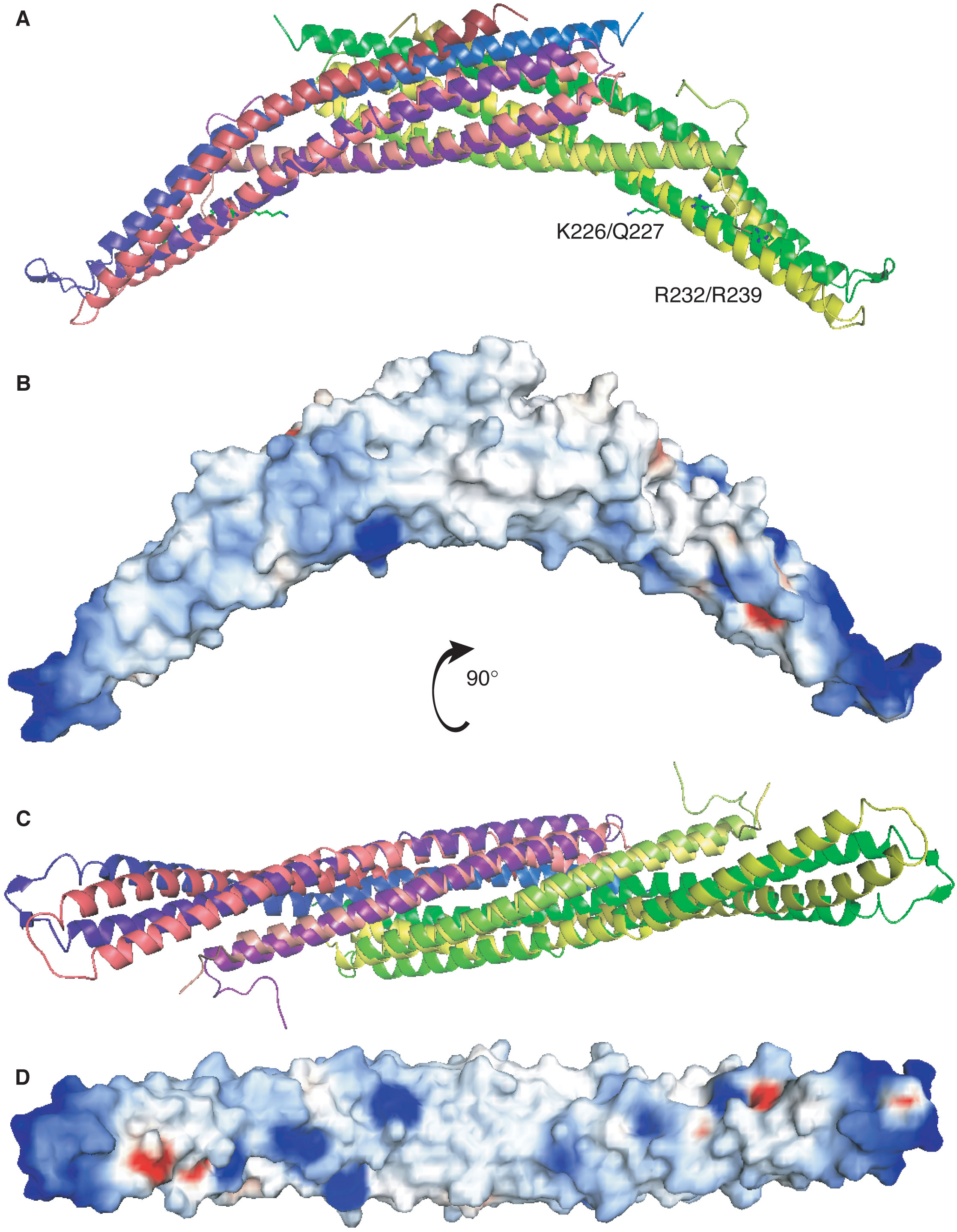
Fig. S5. Overlay of arfaptin and amphiphysin structures
(A) Ribbon representation of the arfaptin C-terminus (in pink and yellow; PDB ID: 1I4D) superimposed on Drosophila amphiphysin BAR (in purple and green; PDB ID: 1URU). The residues of arfaptin that were mutated are shown. (B) Electrostatic surface of arfaptin, as in Figure 1B,D. (C,D) As in (A,B) except the concave surface is shown.
(click on image to see a higher resolution version (716 kb) in a new window)