Lucinda Dawkins
The Perse School for Girls
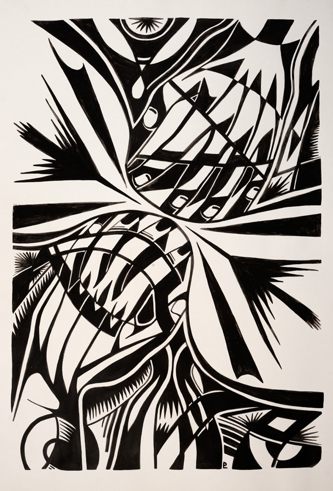
Vesicle Fusion 1
(First image.) An impression of 'Vesicle Fusion' where 2 membranes are depicted in the process of merging.
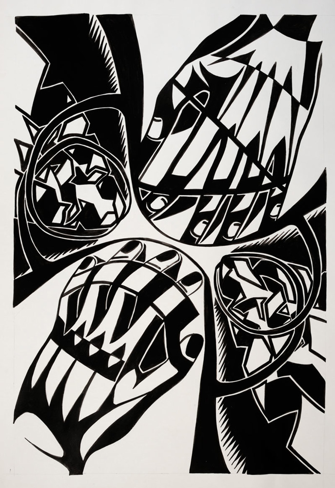
Vesicle Fusion 2
(Second image.) Vesicle fusion driven by SNARE proteins (the helical structures) and synaptotagmin (the membrane bending structures).
Clathrin triskelion
(Third image.) An impression of a clathrin trimer- the structural protein that forms a cage around budding clathrin-coated vesicles.
Budding clathrin-coated vesicle
(Fourth image.) Flexing proteins surround a budding vesicle.
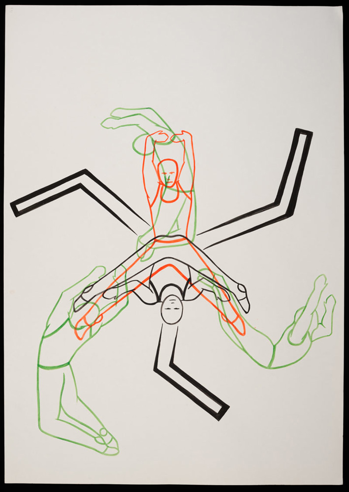
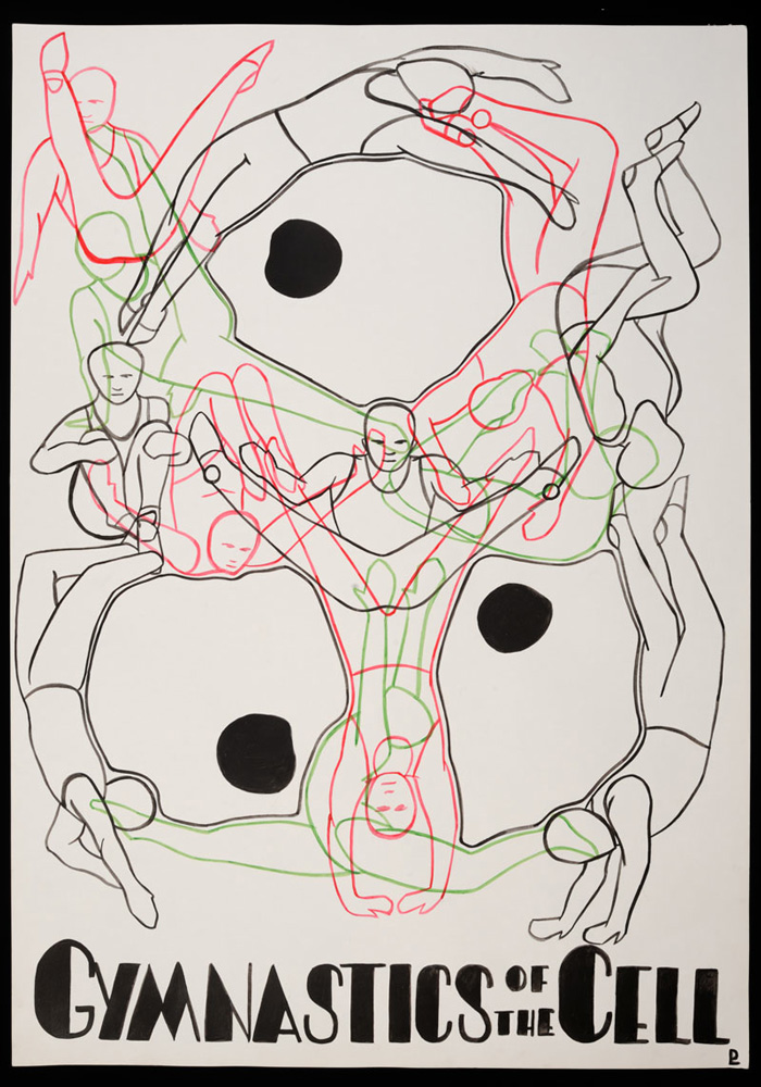
Cell gymnastics 1
(Fifth image.) An impression of the fluidity/flexibility of membranes surrounding cells.
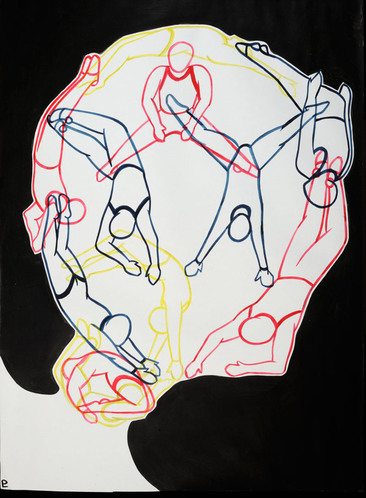
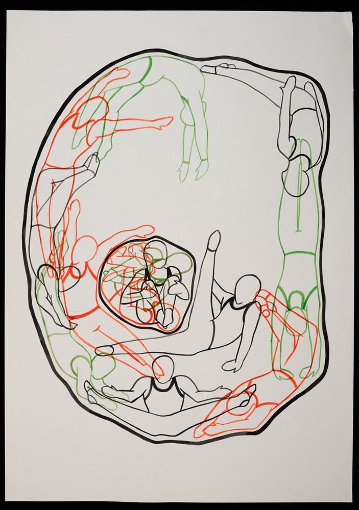
Cell gymnastics 2
(Sixth image.) An impression of the flexibility of membranes in a cell.