Cell Biology of epithelial morphogenesis – understanding dynamic epithelial behaviour
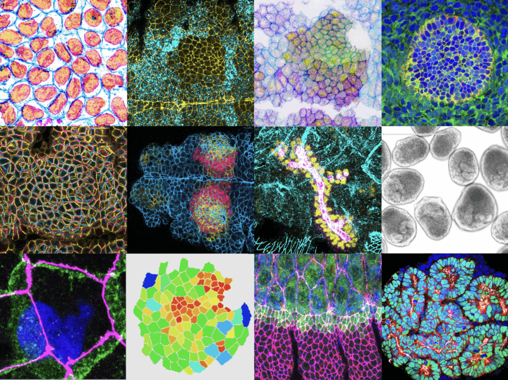
During embryogenesis, organs form by the transformation of simple epithelial layers of cells into complex 3D structures. This remarkable feat is accomplished through the collective and controlled changes in the shapes and arrangements of a large number of cells. The central aim of my group is to understand how genetic programmes drive morphologic changes in individual cells, and how those shape changes are coordinated through cell-cell interactions across an entire epithelium to sculpt a nascent tissue. Because organ shape is critical for organ function, defects in morphogenesis lead to severe diseases including spina bifida or polycystic kidney disease. Thus, understanding the mechanisms that drive faithful organ formation will elucidate a key aspect of metazoan development, help explain the pathogenesis of various developmental diseases, and may lead to new treatment opportunities.
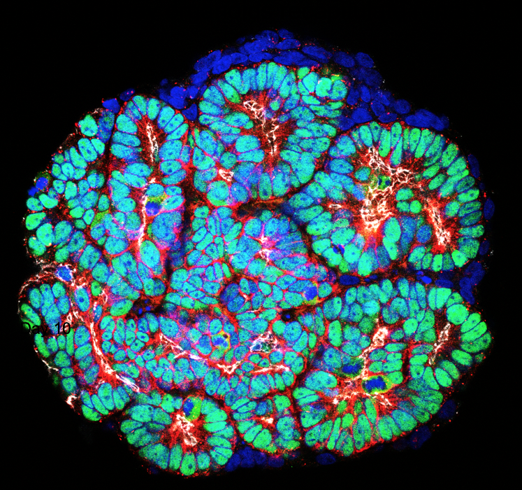
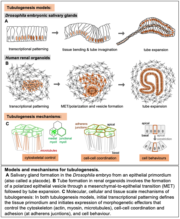
Many of the important organ systems in mammals and invertebrates are tubular in structure, including the intestinal tract, kidney, liver, lung, vasculature, and most glandular organs. Tube formation is thus a critical step in organogenesis, and can occur through a number of mechanisms that include the folding, wrapping, or budding of epithelial sheets. My lab investigates two models of tube morphogenesis. Our major focus has been the salivary glands of the Drosophila embryo , with more recent work also including human renal organoids.
