|
Fibre optics coupled CCD detectors (pre-2000) |
|
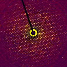
The main emphasis of my work since ~1990 has shifted to the application of semiconductor detectors to electron microscopy with the ultimate aim of being able to completely replace film for all measurements. The first type of detectors, which we investigated were based on low noise CCDs, originally designed for astronomical observations. These devices can only be used in an indirect mode: electrons incident on a phosphor give out light which is optically transmitted to the CCD through fibre-optics. Although the spatial resolution in these devices is restricted due to multiple light scattering within the phosphor grains and at the CCD-fibre optics interface, they are capable of recording electron diffraction.
Publications related to CCD work
- A High Sensitivity Imaging Detector for Electron Microscopy
A.R.Faruqi, H.N.Andrews and R.Henderson
Nucl Instr and Meth A367,408-412, (1995).
- Cooled CCD Camera with Tapered Fibre Optics for Electron Microscopy
A.R.Faruqi and H.N.Andrews
Nucl.Instr. and Meth. A392, 233-236, (1997).
- Electron Diffraction Studies of light-induced conformational changes in the Leu-93 to Ala bacteriorhodopsin mutant
S.Subramanium, A.R.Faruqi, D.Oesterhelt and R.Henderson
Proc. Natl. Acad.Sci. USA Vol 94, 1767-1772, (1997).
- Cooled CCD Detector with Tapered Fibre Optics for Electron Diffraction Patterns
A.R.Faruqi, R.Henderson and S.Subramaniam
Ultramicroscopy, 75, 235-250, (1999).
- Evaluation of Gadolinium oxy-sulphide (P43) phosphor used in CCD detectors for electron microscopy
A.R.Faruqi and G.C.Tyrell
Ultramicroscopy, 76, 69-75, (1999).
- Protein conformational changes in the bacteriorhodopsin photocycle
S.Subramaniam, M.Lindahl, P.Bullough, A.R.Faruqi, J.Tittor, D.Oeterhelt, L.Brown, J.Lanyi and R.Henderson
J.Mol.Biol, 287(1), 145-161, (1999).
- Cooled CCDs for recording data from electron microscopes
A.R.Faruqi
Nucl. Instr. and Meth. A439, 606-610 (2000).
- A Tiled CCD detector with 2 by 2 Array and Tapered Fibre Optics for Electron Microscopy
A.R.Faruqi, H.N.Andrews, D.M.Cattermole and S.Stubbings
Nucl. Instr. and Meth. 477, 137-142, (2002).
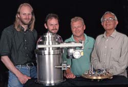 |
The CCD group (from left)
David Cattermole, Howard
Andrews, Steve Stubbings
and Wasi Faruqi. |
|
|
Disclaimer:
These pages are my personal pages. The opinions expressed here are not necessarily those of the MRC Laboratory of Molecular Biology or the Medical Research Council. |
|