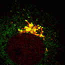
EpsinR WT overexpression in COS cells
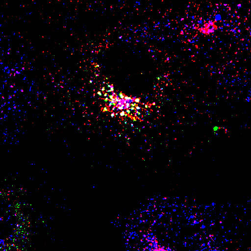 |
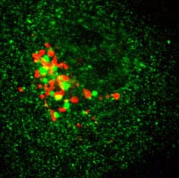
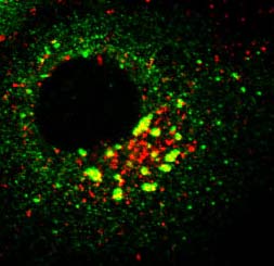
Myc-EpsinR WT with endogenous AP1 and transferrin uptake in COS cells
In the transfected epsinR cell in the image on the left, only the perinuclear puncta significantly co-localise with AP1 and not with internalised transferrin. Endogenous epsinR and overexpressed epsinR do not overlap with overexpressed epsin1 (see link), but there is considerable overlap with TGN markers (see below). With higher levels of epsinR expression there are fewer larger puncta (click here for examples). Click image to view individual channels or proceed to galleries of images below. |
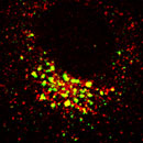
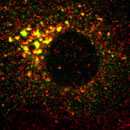
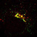
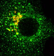
Colocalisation of EpsinR (green) with markers (red), focusing on the perinuclear region.
 |
 |
 |
 |
 |
|
Clathrin |
M6P receptor |
LAMP1 |
||
 |
 |
 |
 |
 |
|
GM130 |
EEA1 |
CD63 |
Blue - Transferrin |
Comparison of epsin1 and epsinR
Myc-epsinR (WT) overexpression and colocalisation with markers
Myc-epsinR D34G+R67L (lipid binding mutant)
Myc-epsinR L10E (proposed lipid conformation mutant)
Myc-epsinR D342R+D422R (clathrin binding mutant)
Myc-epsinR D34G+R67L + D342R+D422R (lipid and clathrin binding mutant)