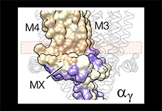
Sub-membrane helix MX
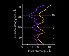
Several decades ago, electrophysiologists working with normal and denervated frog muscle fibres discovered that there are two populations of ACh receptors: junctional receptors, i.e. those located within the neuromuscular junction; and extra-junctional receptors, present in more distant regions of the plasma membrane. The junctional receptors exhibited more rapid gating kinetics and were 2-3 times more conductive than those in extra-junctional regions.
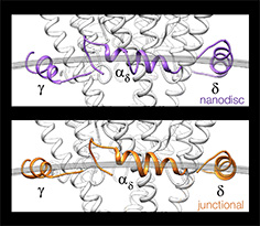
The cell membrane in the junctional region has a uniquely asymmetric and ordered structure, due to a high concentration of cholesterol. Cryo-EM studies show that this special lipid environment influences the transmembrane (TM) folding of the receptor. The sub-membrane helices MX may adopt poses similar to those in nanodisc-solved structures in the lipid-disordered extra-junctional regions. But at the neuromuscular junction the helices engage with the cholesterol-ordered lipids, forming a coplanar arrangement. Realignment of MX to the flat bilayer surface causes the coupled TM helices to splay apart, creating the fast-acting form of the receptor that operates at the synapse.
Key publication:
Unwin, N. Influence of lipid bilayer on the structure of the muscle-type nicotinic acetylcholine receptor. Proc. Natl. Acad. Sci. USA 121, e2319913121 (2024). (pdf)