Welcome to the Scheres lab
We are intrigued by how proteins work. The genetic code determines the amino acid sequence of proteins, which in turn determines their 3D-structure. The precise 3D-arrangement of different types of atoms inside proteins allows them to perform the many types of functions that are needed to keep the cell alive. Therefore, visualising the 3D-structures of proteins is a powerful way to find out more about how they work. Research in our lab focuses on different aspects of protein structure determination.
One line of work focuses on the development of image processing methods to determine protein structures from electron cryo-microscopy (cryo-EM) images. We implement these methods in our open-source computer program RELION. This program is used in many cryo-EM labs all over the world and has been used to solve numerous protein structures.
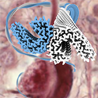
In another line of research, we use cryo-EM to solve our own protein structures. However, instead of solving structures of proteins in their functional state, we focus on protein structures that are related to diseases: amyloid filaments. For this purpose, we needed to expand our image processing methods to allow atomic-resolution structure determination of amyloids. We now apply these methods to study filaments that are extracted from the brains of diceased individuals with different types of neurodegenerative diseases, like Tau filaments from Alzheimer's (image above) and chronic traumatic encephalopathy (CTE).