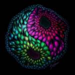
An artist’s rendition of the diverse cells within an organoid and the balance between different brain regions. Cover image for Renner M, Lancaster MA, et al. EMBO J. 2017. Credit: Beata Mierzwa.
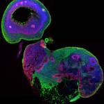
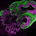
A section through a whole organoid stained for neurons in green and neural stem cells in red. All cell nuclei are stained in blue.
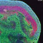
A section through a single lobe of cortical tissue within an organoid. Neurons are in green, neural stem cells in red, and all nuclei are stained in blue.
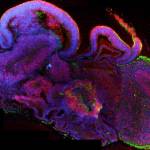
A section through a whole organoid stained for neurons in green and neural stem cells in red. All cell nuclei are stained in blue
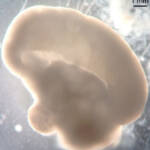
A whole organoid imaged in bright field. Note the large lobe of cortical tissue and the overall large size of the organoid reaching approximately 1 cm in length.
