 |
 |
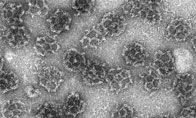
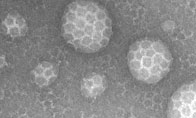
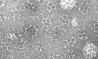
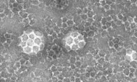
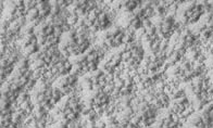
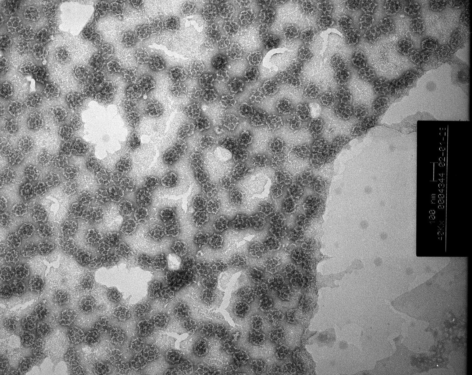
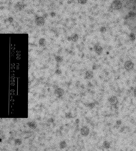
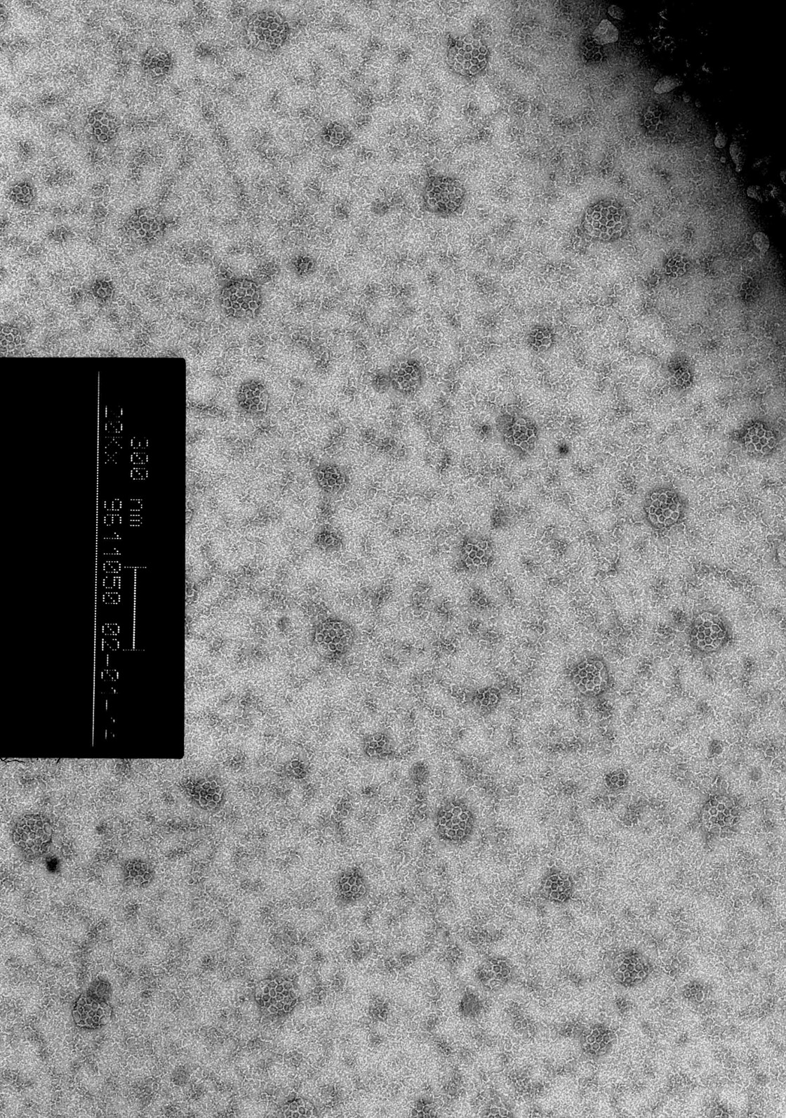
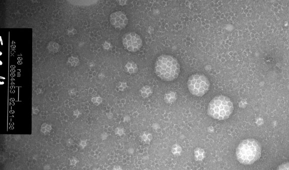
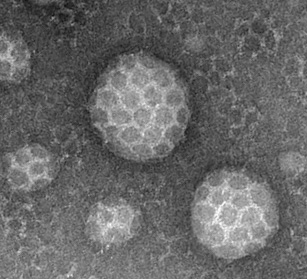
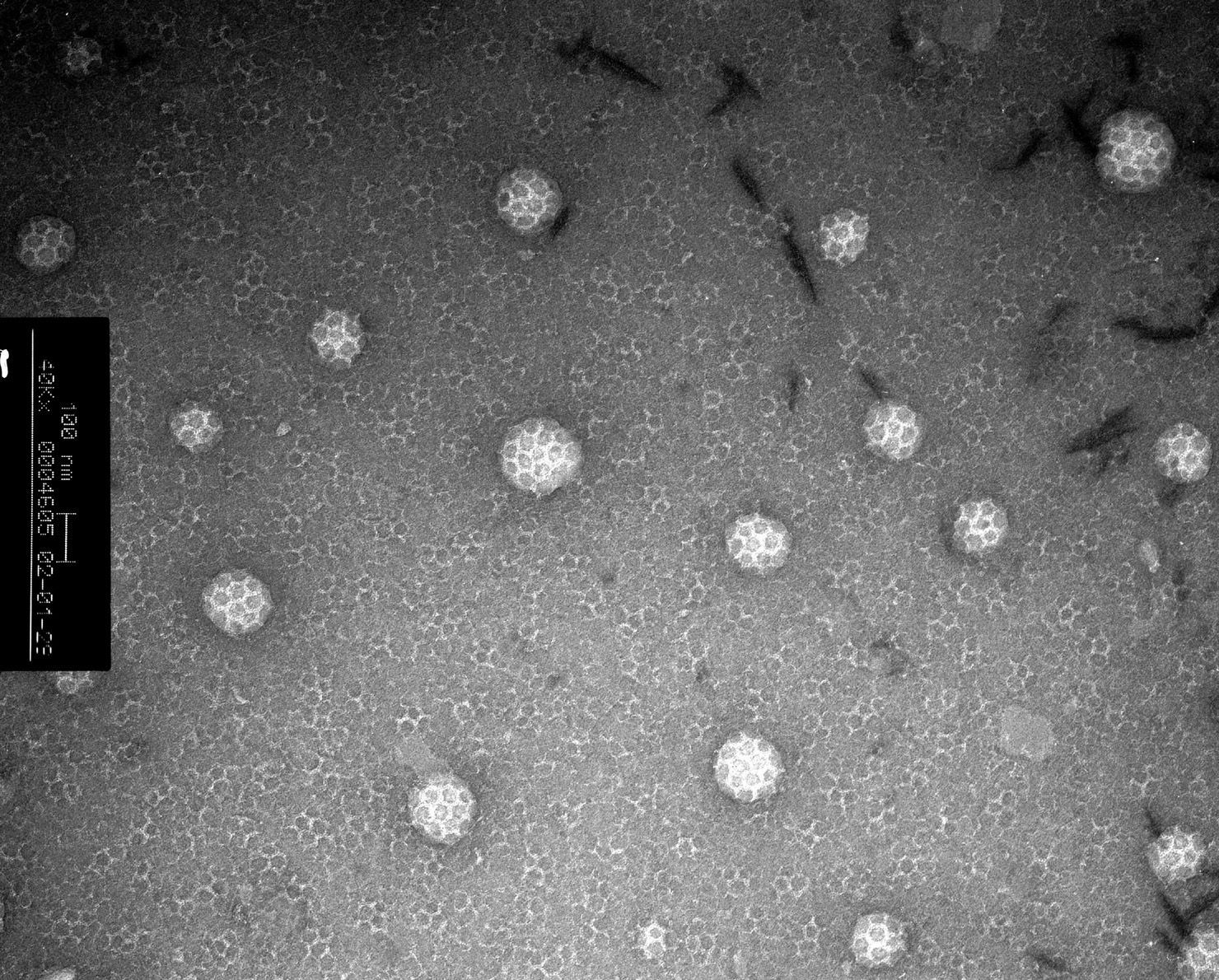
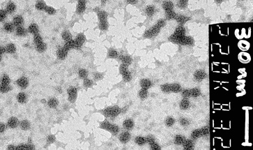
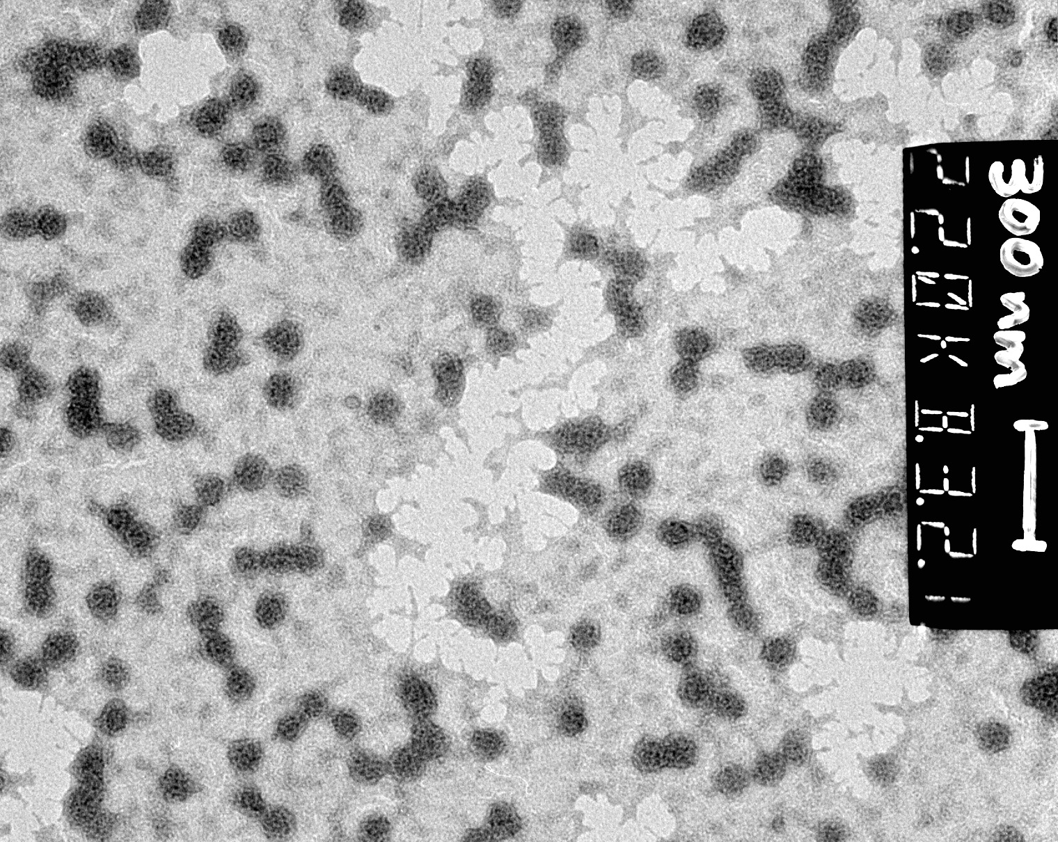
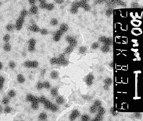
AP180 + Clathrin
|
|
Small image (141 kb) Small image (53 kb)
 |
 |
 |
| Small stereo image (48 kb) | Small image (87 kb) | Small image (61 kb) |
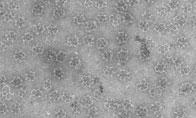
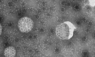
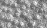
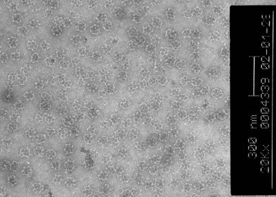
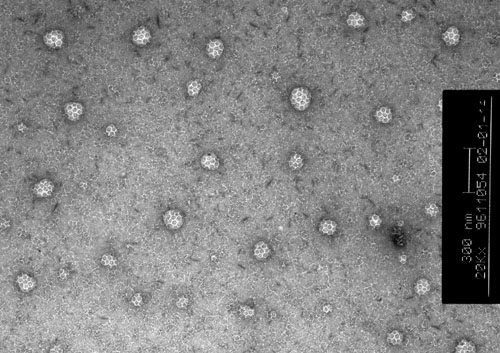
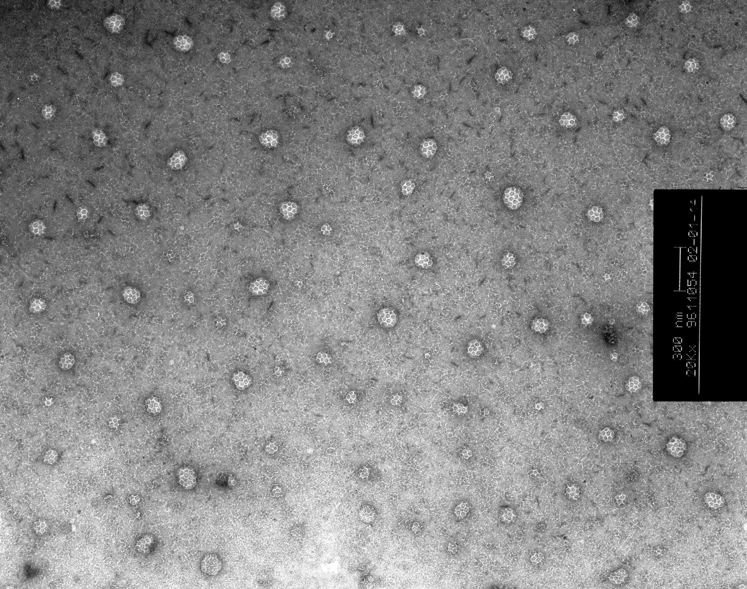
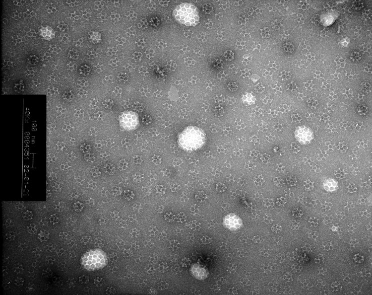
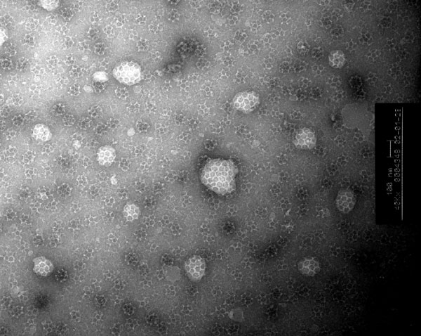
AP180 + Epsin + Clathrin
 |
 |
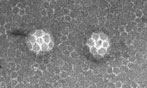 |
| Small image (46 kb) | Small image (68 kb) | Small image (74 kb) |
 |
 |
 |
| Small image (76 kb) |
|
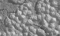
Small image (131 kb) |
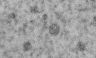
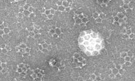
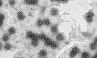
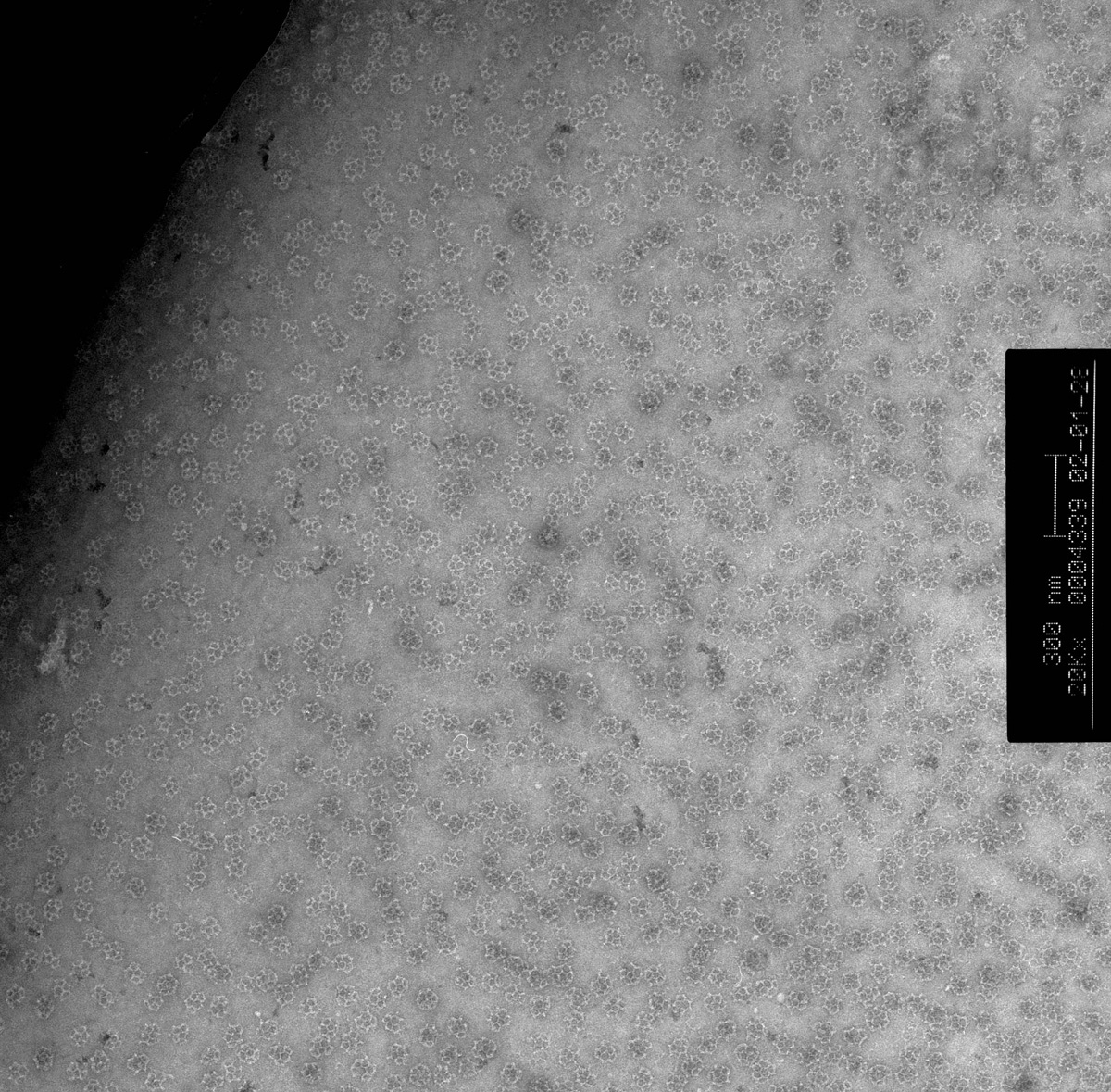
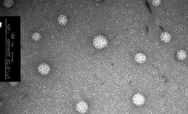
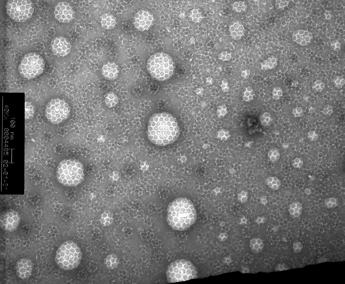
AP180 + AP2 + Clathrin
 |
 |
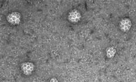
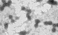
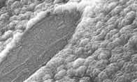
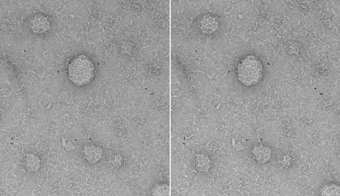
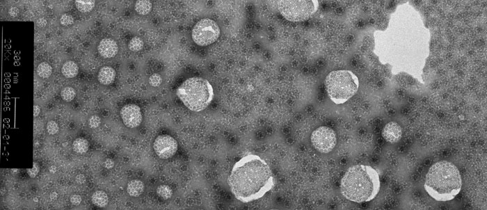
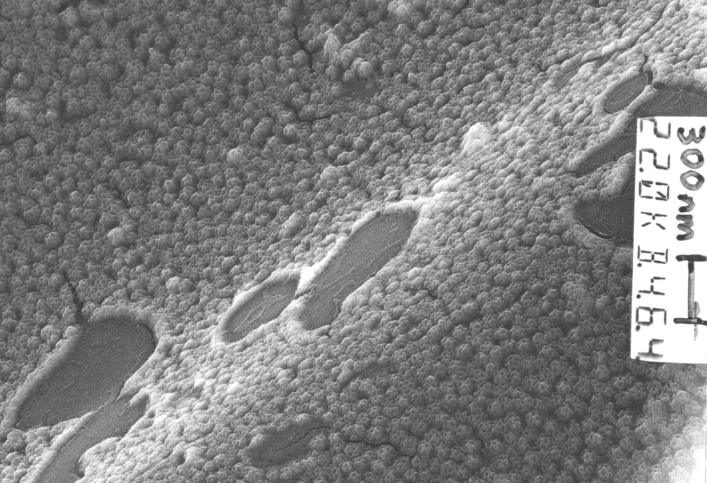
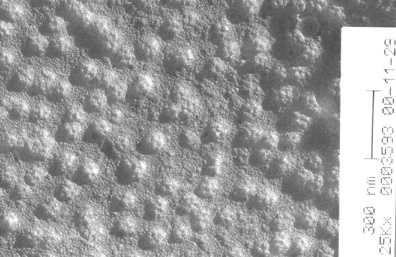
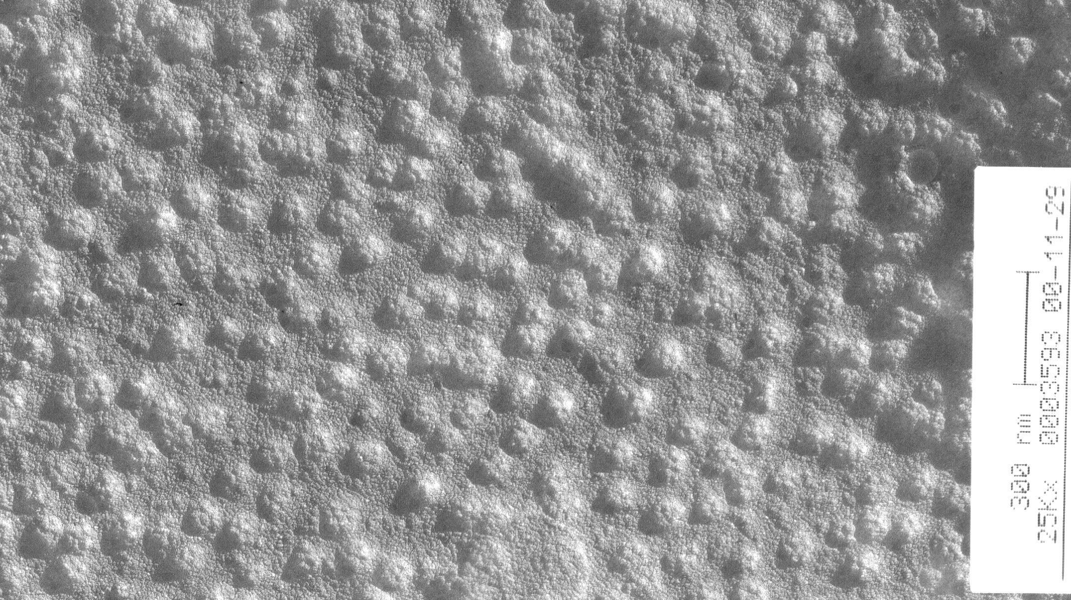
AP180 + Clathrin, platinum shadowed
 |
 |
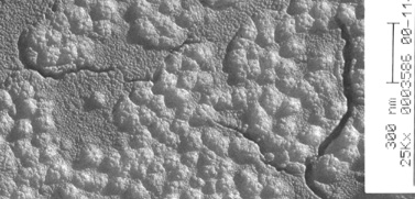
AP180 + AP2 + Clathrin, platinum shadowed
 |
 |
Other EMs (limited access)