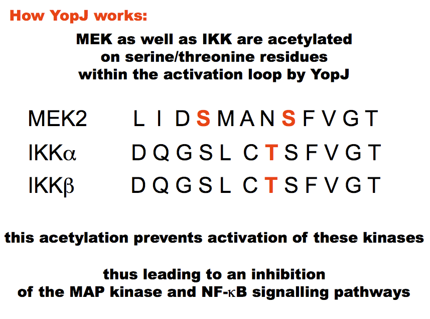
Yersinia bacteria (the causative agents of plague and other gastrointestinal disorders) inject a mixture of toxins including YopJ into mammalian host cells. This prevents their engulfment by macrophages and YopJ suppresses the innate immune responses. The key enzymes targeted by YopJ are the MAPKKs of the MAP kinase signalling pathway and IKKs of the NFkB pathway.
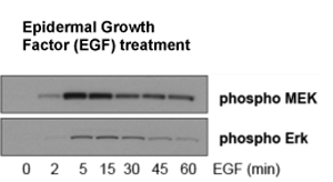
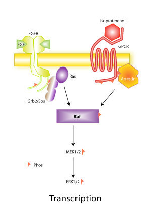
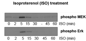
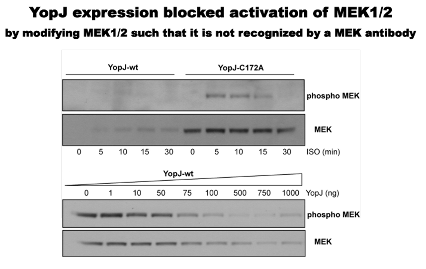
The MAPK pathway can be activated by different stimuli including Epidermal Growth Factor (EGF) acting on receptor tyrosine kinases, and by isoproterenol acting on G-protein coupled receptors (GPCRs). The activation on receptor binding is rapid and results in the activation of a series of kinases collectively known as the MAP kinase cascade. The activation and inactivation kinetics can be followed by the use of phospho-specific antibodies against members of the pathway (for example MEK and Erk shown here).
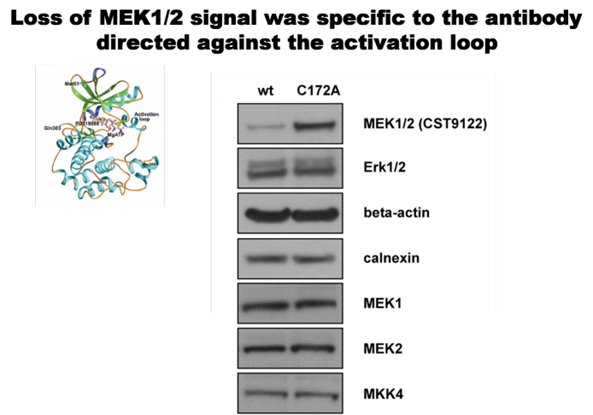
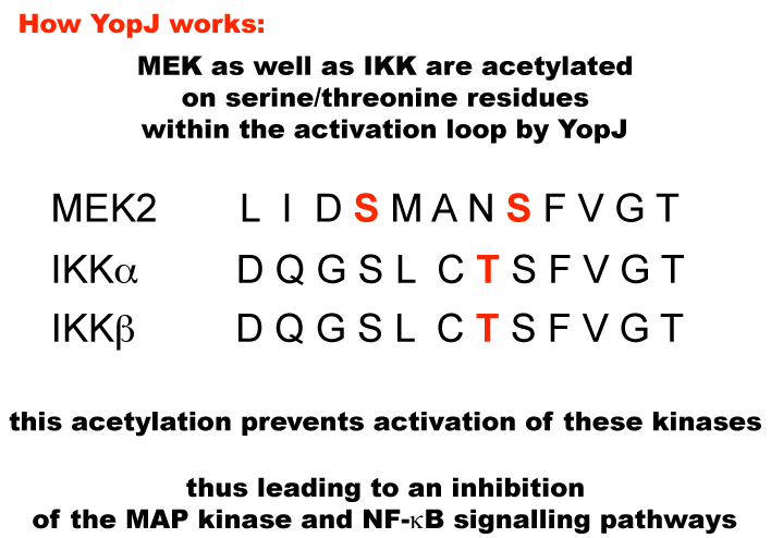
We found that an antibody recognizing the activation loop of MEK would no longer recognize its target due to acetylation of the loop.
Antibodies directed against other regions of this kinase could still recognize YopJ-modified MEK.
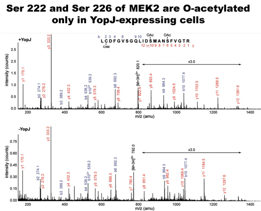
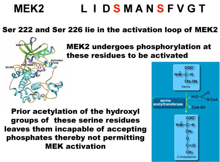
The above results suggested that YopJ was modifying the activation loop and by Mass spectrometry we identified the modified residues to be serine 222 and serine 226 in MEK2. These MEK samples were prepared from cells transfected with MEK-His6 and YopJ.
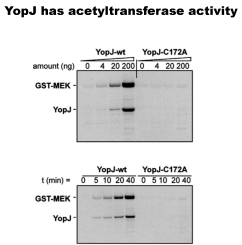
In vitro we could demonstrate that YopJ itself has the acetyltransferase activity, and a mutant C172A was inactive. We also could demonstrate that YopJ is autoacetylated.
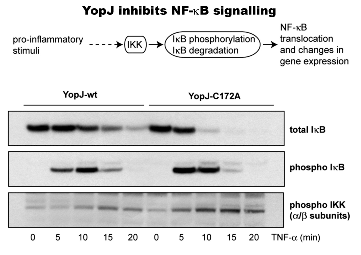
Pro-inflamatory stimuli ultimately result in the activation of the IKK IkappaKinase complex. The results in the phosphorylation of IkB (an inhibitor of NFkappaB). This phosphorylation results in the ubiquitination and subsequent proteosomal degradation of IkB. This free NFkB to translocate to the nucleus and activate the transcription of gene with an NFkB regulatory element.
The panel shows that YopJ-wt inhibits IkB degradation (top panel) as well as phosphorylation (middle panel). This is due to inhibition of activation (by phosphorylation) of IKKs (bottom panel).
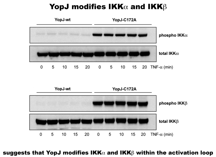
The autophosphorylation of both IKKalpha and IKKbeta is blocked by overexpression of YopJ-wt in cells (and C172A mutant is ineffective). This suggests that modification by YopJ prevents the activation of IKKs.
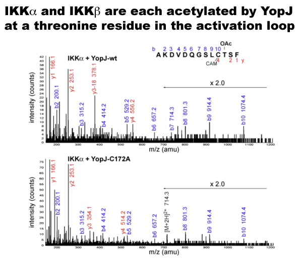
IKKalpha and IKKbeta immunoprecipitated from cells were shown to be acetylated by expressed wild-type YopJ and not by the C172A mutant.
Our publications on YopJ Mittal et al PNAS 2006
Mittal et al JBC 2010