
About Us
Microscopes4Schools is a hands-on science outreach activity for primary school children run by scientists from the MRC Laboratory of Molecular Biology.
Microscopes4Schools began with a casual chat about how great it would be to take microscopes to primary schools and show cells to children. As cell biologists, we couldn't think of anything more exciting for children to do in the classroom – it’s probably not an overstatement to say that all biologists cherish their memories of the first time they looked down a microscope. Discovering the microscopic world is an amazing experience and we wanted to share it with as many children as possible.
The founders of M4S, Simon and Isabel, put their ideas onto paper and came up with a project for an outreach activity for primary schools. They were lucky to have generous financial support of the MRC and the Lister Institute for Preventive Medicine, who were equally excited about taking cell biology and microscopes to the classroom.
What kind of microscope do the schools receive?
After some research, they found some affordable educational microscopes of very good quality. It is still astonishing how impressive the images are that the children capture – many images are of a similar quality to those taken with much more expensive research microscopes.
The excitement and interest of the children during our school visits surpassed all our expectations.
Will the children understand?
We had feared the children were too young to be able to look down a microscope, but they can do it very easily. We weren't sure they would grasp the abstract concept of cell if they had not heard of it before . However, this was also not the case.
Now, the competition has adapted throughout the COVID pandemic to include a virtual session with scientists rather than an in-person visit (which we aim to resume when possible).
Having seen the children's enthusiasm and keen interest of the teachers to use microscopes in their classroom, this website supports the project and its competition with all the necessary information. We hope this allows more teachers and families to learn about which microscopes to buy, how to use them, and what fun biological samples are easy to access.
We hope that teachers, parents and children will be encouraged by this website to use a microscope and that this will help spark their curiosity in the living world.
Who we are
Simon Bullock
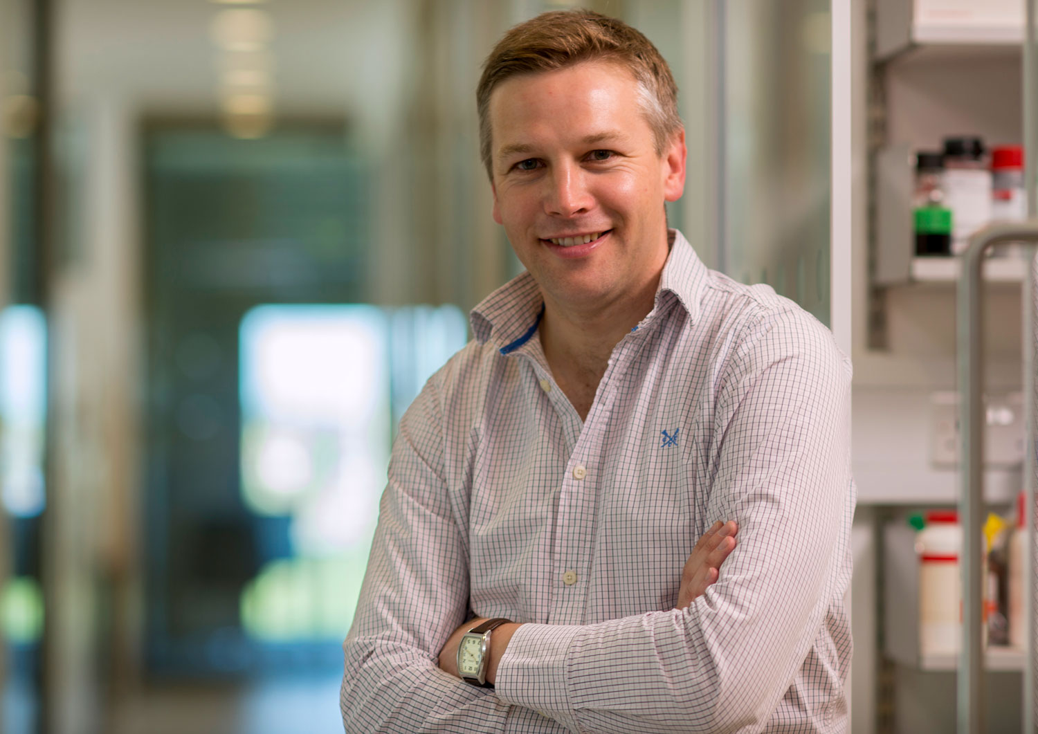 Simple experiments with microscopes played key roles in shaping my career choices. Seeing the beating heart of a Daphnia down a microscope at school when I was 16 played a significant part in my decision to study Biology at university.
My long-standing interest in cell dynamics can be traced back to watching cell divisions in frog embryos in a university practical. My experiences made me appreciate that seeing the microscopic world with your own eyes can be a great way to spark an interest in science, and that it would be great to give younger children that opportunity as well.
Being involved with Microscopes4Schools has been extremely rewarding and stimulating. For this reason, I would strongly encourage other scientists to set up their own outreach activities.
Jonathan Howe
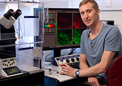 My interest in microscopy began during my biology lessons at school. I remember how cool it was to see cells for the first time when we prepared and stained onion cell samples. Clearly this also had a profound effect on several of my classmates as there was a lot of crying going on in the classroom that day!
During my time at university, I was introduced to the beautiful world of fluorescence microscopy. I have a vivid memory of observing fluorescent vesicles (compartments of the cell) whizzing around and realising that this happened so quickly it was only possible to appreciate by eye with the help of a microscope.
Currently, I work in the Light Microscopy facility at the LMB and help scientists to study a wide variety of biological processes on our microscopes. These range from cell division to protein transportation. I find it extremely important and rewarding to participate in public engagement activities, such as the Microscopes4Schools initiative. Seeing the excitement in the children reminds me why I pursued a career in science.
Isabel Torres
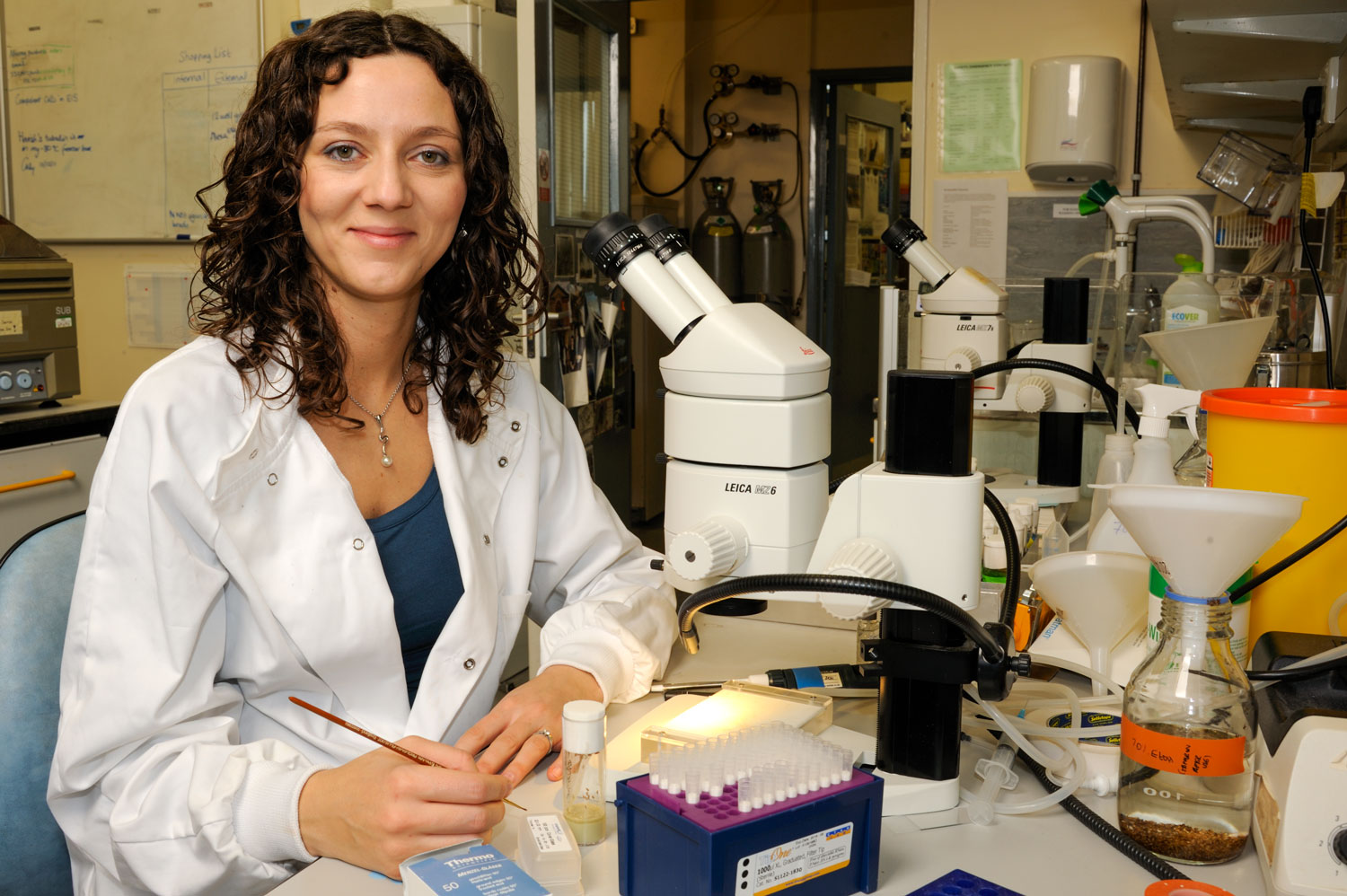 Isabel was a co-founder of M4S with Simon and was at the LMB between 2008-2012. Her curiosity led her to become a scientist and she was driven by her interest in understanding how things work. At school, Isabel liked all sciences, but was most fascinated with Biology. Isabel has always found that explaining science to the public is incredibly rewarding and lots of fun.
Meet the Scientists
In addition to lending schools a microscope for the competition, the M4S Competition provides an online session for each school during the two-week competition slot.
These sessions are hosted by LMB scientists who give their time and help provide pupils with the chance to see what incredible work takes place in our labs as well as asking their own questions about any aspect of science, including careers.
2022 Meet the Scientists
 Magda works on brain development and loves to look after her cells. She is fascinated by nature and believes the closer you can look at natural objects with the help of microscopes, the more beautiful nature becomes. Madga grows miniature brain models called organoids to help her understand how signalling occurs in neurons.
 Lorena aims to understand how dendritic cells can tell the difference between healthy and unhealthy cells in the body. Dendritic cells are a type of immune cell which help to organise an immune response to stop viruses, bacteria and cancer cells from growing in the body. Her research focuses on how cancer cells avoid being detected by dendritic cells, and how we can make these cancer cells more visible to dendritic cells so they can be removed by the body’s immune system.
 Dan is an Australian who is working in Cambridge. Dan is a neurobiology researcher working on understanding the brains in flies. When he’s not doing science in his spare time, Dan likes to sing and go camping.
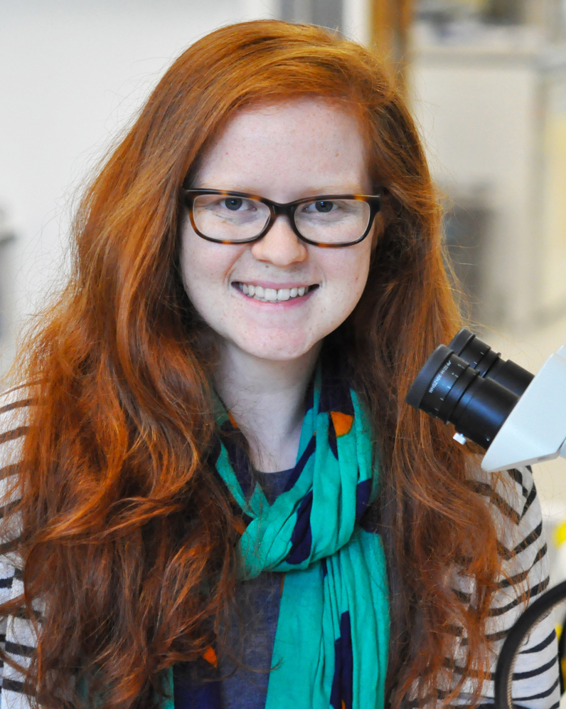
 Elyse works on cell division and what regulates that in cells. She recently finished her PhD and has stayed on in the lab. Elyse was inspired by her grandfather who was also a scientist and remembers looking down the microscope at cheeses as a child!
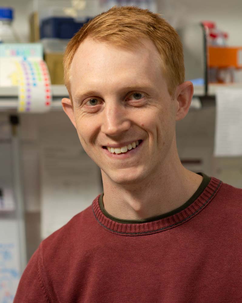
 Alex’s research focuses on how things are moved around cells and how this process is regulated by the motor protein dynein. Alex uses light microscopy to look at how cells transport smaller compartments called mitochondria, the power houses of cells. Now, he combines the techniques he learnt to better understand this process in human neurons.
 Annabel works on fruit flies and uses various methods to help understand how a single sheet of cells can form tubes. Annabel often uses microscopes in the work in the lab to help see these structures more clearly and with fluorescent dyes that turn the cells different colours.
 Nathan works largely with electron microscopy and imaging cells to understand cellular rhythms, signalling and regulation of cell energy.
 Sam is working on understanding the origins of life. He tries to work out how biological molecules first appeared on the early Earth.
 Alice is a post-doctoral researcher (so has studied science at university and then did a PhD degree in the field of Biochemistry). During her PhD studies, Alice designed experiments to investigate how eukaryotic cells can take in nutrients from outside of the cell and then deliver them to the correct place inside the cell. Alice is interested in looking at the cell membrane of archaea as in archaea, the cell membrane is a thin layer that separates the cell from the outside world.
 Aly is interested in how embryonic stem cells are able to form the different cell types of the body. To do this, he uses a fluorescent microscope to watch stem cells as they grow and see all the kinds of cells that they make. He also takes lots of 3D images of embryos to understand how this happens in actual living organisms.
 Andrew investigates the body clock - the way our body tells us it’s time to stay awake in the daytime and get sleepy at night time. He’s also really interested in why some animals do the opposite and stay awake at night time, like mice, hedgehogs, and other animals.
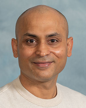 Bilal loves taking images of cellular components using a powerful 3m tall electron microscope which can make images over a hundred-thousand times bigger than the object! These images can be used to understand the structure of molecules and apply this to drug discovery in science, building on our understanding of cellular machines at work inside the cell. Bilal has studied in Canada, Singapore and now moved to Cambridge, UK to help other scientists master the techniques he uses. He loves to share his knowledge so the M4S competition was a great opportunity to do this with curious young minds.
 Florence is interested in the microscopic machines that move things along tracks inside our cells. Her PhD investigated how these machines work in neurons. She often uses microscopes to see these machines moving, such as inside the wings of fruit flies!
 Kat uses electron microscopes in her PhD work on transport compartments in the cell. She is also interested in the structure of viruses, like the coronavirus. She uses electrons, not light, to be able to zoom in on the tiniest of details and understand how things are built.
 Katherine is a first year PhD student in the Cell Biology division at the LMB. Before that, Katherine studied Biomedical Science at The University of Sheffield. She uses light microscopy to understand a compartment within cells called the Golgi apparatus. Once proteins have been made from the cell’s genetic code (the recipe for proteins), the Golgi is responsible for adding additional modifications (like icing on a cake) and making sure that once these modifications have been added, that the proteins are delivered to the right parts of cell (like a cake delivery service) and this helps cells to function.
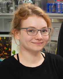
 Ella is a second year PhD student in the Röper lab which is part of the Cell Biology division. Her lab is interested in how during development, very early changes in transcription factors (which regulate what genes are currently switched on), change the shape of the developing organism. She works with kidney organoids (mini kidneys in a dish) and tries to work out what genes and mechanisms control the formation of tubes as the kidney grows.
 Stephan works with scientists who want to understand how structures called amyloid filaments form in the brain. These filaments are involved in diseases like Alzheimer’s disease. When this happens, it can cause cells in the brain called neurones to break down and means people lose functions like memory. Stephan also focuses on who may be at risk of these diseases to help scientists make better medicines.
 Kendra uses a very powerful microscope to study proteins that help cells detect when they have been infected with a virus and activate antiviral defence mechanisms. By studying the structure of these proteins, the lab hopes to understand how they work and what happens when they don’t function as they should. Kendra is also very interested in understanding the structure of viral proteins and how we can use this knowledge to prevent or treat viral diseases.
We’re currently recruiting LMB scientists for these slots so please get in touch.