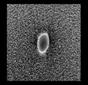
Electrostatic phase plate
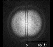
A standard way to bring out the shape or surface details of a protein molecule in the electron microscope is to embed it in a heavy metal stain. But only the stain is well contrasted, while the protein underneath remains invisible – making internal features impossible to visualise. One attempt to overcome this problem involved development of an electrostatic phase plate which highlights the encased protein, while diminishing contrast from the stain. The phase plate has a thin (0.3μm dia.) gold-coated spider's thread spanning the objective aperture, which forms a stable charge in the region of the central direct beam. The electric field created is cylindrical locally, but then becomes more spherical, producing at higher angles a uniform phase shift that delivers ‘bright’ phase contrast. This is the opposite to the ‘dark’ contrast achieved by under-focusing and, together with attenuation of the direct beam, largely cancels out the dark appearance of the stain.
Key publications:
Unwin, P.N.T. Phase contrast and interference microscopy with the electron microscope. Phil. Trans. Roy. Soc. Lond. B. 261, 95-104 (1971). (pdf)
Unwin, P.N.T. Electron microscopy of biological specimens by means of an electrostatic phase plate. Proc. R. Soc. Lond. A. 329, 327-359 (1972). (pdf)
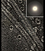
Unwin, P.N.T. and Klug, A. Electron microscopy of the stacked disk aggregate of tobacco mosaic virus protein. 1. Three-dimensional image reconstruction. J. Mol. Biol. 87, 641-656 (1974). (pdf)