Low-dose microscopy
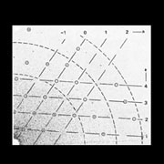
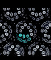
Another imaging approach, that led to much more informative pictures, was to eliminate the stain altogether and image the radiation-sensitive protein directly, using a low electron dose. Although the weak dose needed to keep structure intact meant that features of individual molecules were very noisy, it was nevertheless possible to build up a detailed image by averaging over the many identical copies comprising a crystalline array. The averaging reinforces genuine features common to each molecule, while statistical fluctuations associated with the low numbers of electrons are smeared out.
In analysing bacteriorhodopsin, glucose was used to substitute for the water that is lost to the microscope vacuum. Plunge-freezing to create a thin film of amorphous ice, and microscopy using a cold stage, is now the method of choice for retaining specimens in a hydrated state.
Key publications:
Unwin, P.N.T. and Henderson, R. Molecular structure determination by electron microscopy of unstained crystalline specimens. J. Mol. Biol. 94, 425-440 (1975). (pdf)
Unwin, P.N.T. Beef liver catalase structure: interpretation of electron micrographs. J. Mol. Biol. 98, 235-242 (1975). (pdf)
Taylor, K.A., Milligan, R.A., Raeburn, C. and Unwin, P.N.T. A cold stage for the Philips EM300 electron microscope. Ultramicroscopy 13, 185-190 (1984). (pdf)
Toyoshima, C. and Unwin, N. Contrast transfer for frozen-hydrated specimens: determination from pairs of defocused images. Ultramicroscopy 25, 279-292 (1988). (pdf)