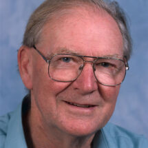
John Finch, a superb crystallographer and later electron microscopist, who joined the LMB when it was founded in 1962, died on Tuesday 5th December, after a short illness. His contributions to the structures of viruses and nucleosomes, and elucidation of the mechanism of negative staining EM, were central to many of the molecular structures solved at the LMB.
John was born on 28th February 1930 in London and attended local schools before winning a scholarship to King’s College London, where he studied physics, graduating in 1955. He then responded to an advert by Rosalind Franklin, at Birkbeck College, for an assistant to help with the work on the structure of viruses. He joined as a technician but very soon afterwards started his PhD.
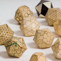
John began with X-ray studies of Tobacco Mosaic Virus (TMV), before deciding to concentrate his studies on small spherical viruses, in particular Turnip Yellow Mosaic Virus (TYMV). JD Bernal’s group, also at Birkbeck, had already obtained powder photographs of small spherical viruses, such as the Tomato Bushy Stunt Virus and TYMV and showed that the symmetry was cubic. From the size of the unit cell and from the size of the virus particle they found that there was only one virus particle per unit cell, roughly speaking, so the virus particle would have to be cubic too, and composed of identical subunits. John was interested in exploring this further. Through X-ray diffraction methods, he found that TYMV displayed icosahedral symmetry and found the same strong evidence for icosahedral symmetry in the diffraction patterns of other small spherical viruses, such as Polio. In 1958, following the untimely death of Rosalind Franklin, her colleague and friend, Aaron Klug took over the group. John completed his PhD in 1959 on ‘X-ray diffraction studies on viruses’.
By this time, Aaron had accepted an invitation for the group to join MRC funded scientists Max Perutz and John Kendrew at a new lab being planned in Cambridge. The LMB opened in February 1962 and the group from Birkbeck moved in, and came to be known as the ‘virus group’.

The move to Cambridge coincided with a move away from X-ray diffraction and John began using electron microscopy (EM). His mastering of this technique would yield significant results. John was the first to show the substructure of TMV directly by electron microscopy, and later determined its hand by methods that would then become standard. Either alone, or sometimes in collaboration with Aaron, he solved the overall structure of many different spherical viruses including Tomato Bushy Stunt, R17, human wart and polyoma, and showed that they conformed to the theoretical predictions of both Aaron and Don Caspar, a former colleague and collaborator. To accomplish this, John elucidated the mechanism of negative staining, then widely used but little understood. His tilting experiments showed conclusively that the EM images represented a superposition of both sides of the particle, not simply a footprint or cast of one side, as was widely believed at the time. This work set the standard for rigorous fine-structure microscopy and paved the way for 3D image reconstructions. John was the first to apply 3D image reconstruction to the paramyxoviruses, and also determined the surface lattices of other structures such as bacterial flagella and fibres of sickle cell haemoglobin. His studies on the protein disk of TMV gave essential insights into the way the disk transforms to the helical state of the virus and laid the basis for the eventual solution of the disk structure to 2.8 Å resolution.
The ‘virus group’ then diversified to look at protein-nucleic acid structures. John made decisive contributions to work on the structure of chromatin, using EM to show that the products of chromatin split enzymatically consisted of single, double, triple beads etc., showing an exact correspondence between chemical and physical structure. He also showed that the basic 100Å fibre of chromatin could be coiled in to a superhelical structure to form a 300Å fibre, corresponding to the state of bulk chromatin in vivo. John was also largely responsible for the X-ray and EM studies of crystals of nucleosomes leading to evidence of the arrangement of the DNA and orientation of the DNA superhelix and of the histone core. Throughout, John’s combined work by EM and X-ray has led to a steady succession of solved structures. He was made a Member of EMBO in 1978 and a Fellow of the Royal Society in 1982.

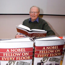
John retired from research in 1995, but stayed on at the LMB as a ‘retired worker’. In 1997, at the request of Richard Henderson, who had recently taken on the role of LMB Director, John began work on writing a book about the history of the LMB. Richard’s initial idea was for the book to follow what happened to the people and groups who made up the intake into the LMB when it was opened in 1962: a ‘family history’ of the laboratory. John put the family in context with the LMB’s history, the pre-LMB history and included other groups and people who had joined the LMB later. In 2008, ‘A Nobel Fellow on Every Floor’ was published by the LMB. The book captures and illuminates the excitement of the inception and development of molecular biology, through the work of the LMB, and remains a key reference point for both staff and alumni.
John Finch was a first-class experimental crystallographer with wide experience of many different systems and the ability to handle difficult crystals. He was a pioneer of fine structure electron microscopy and set the standards for others in the field. He will be remembered with fondness and admiration.