
Dyneins are a family of motor proteins that move along microtubules to transport various important cargos, including proteins and RNAs, to different parts of the cell and are crucial to correct cell function. Gradually, the structure of various components of dynein have been revealed. Now work by Andrew Carter’s group in the LMB’s Structural Studies Division, in collaboration with Alexander Bird’s group at the Max Planck Institute Dortmund, has determined the 3D structure of the whole human cytoplasmic dynein-1 in an inhibited form and shown how it is activated.
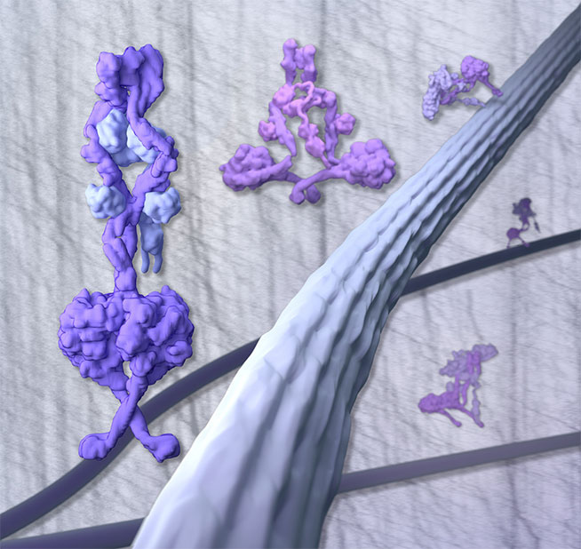
Cytoplasmic dynein-1 binds dynactin, the protein complex that activates it, and cargo adaptor proteins to form a transport machine capable of long distance processive movement along microtubules. Until now it was unclear why dynein-1 moves poorly on its own or how it is activated by dynactin. Using a combination of electron cryo-microscopy (cryo-EM), in vitro assays and cell assays, Andrew’s group have determined the complete 1.4 MDa human dynein-1 complex in an inhibited state, known as the phi-particle. This structure shows how the two motor domains in dynein dimerise and explains how this inhibits dynein.
Disrupting motor dimerisation with structure-based mutagenesis drives dynein-1 into an open form with higher affinity for both microtubules and dynactin. They found that the open form is surprisingly still inhibited for movement and that dynactin relieves this by reorienting the motor domains to interact correctly with microtubules. This model explains how dynactin binding to the dynein-1 tail directly stimulates its motor activity.
This work also showed that disrupting the phi-particle changes dynein’s location and leads to defects in cell division. A number of human mutations that cause malformation of cortical development (MCD) map to the interface between dynein motor domains and so may work by disrupting the phi-particle. Although MCD mutations are very rare, five different mutations map to this same interface. Dynein is also known to be involved with many diseases, for example viral infections, and a better understanding of how dynein works might lead to new ideas for the treatment of these diseases.
The work was funded by the MRC and a Wellcome Trust New Investigator Award.