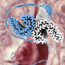
Alzheimer’s disease, the most common neurodegenerative disease, is characterised by the formation of filamentous Tau protein inside nerve cells and amyloid-beta peptides outside cells. Despite more than three decades of research into Tau filaments from a range of different neurodegenerative diseases, atomic structures were still lacking. Now, research by the groups of Sjors Scheres in the LMB’s Structural Studies Division and Michel Goedert, in the Neurobiology Division, in collaboration with Bernardino Ghetti at the Indiana University School of Medicine, has for the first time revealed the atomic structures of Tau filaments.
This groundbreaking research involved the extraction of Tau filaments by Ben Falcon from the brain of a patient who had died with Alzheimer’s disease. These filaments were then imaged using electron cryo-microscopy (cryo-EM) by Anthony Fitzpatrick to determine the atomic structures of their ordered core. Imaging filaments that were extracted from diseased brain is significant, as previous work depended on samples that were assembled in the lab. Because amyloid structures can form in many different ways, it remained unclear how closely such structures resembled those in human disease. However, Tau filaments are extremely ‘smooth’, so it is difficult to superimpose images at the atomic level by cryo-EM. The development of new software by Shaoda He and Sjors was crucial for calculating the structures of the filaments to sufficient detail to deduce the arrangement of the atoms inside them.
The resulting atomic structures teach us which parts of Tau form the seed for aggregation. This is relevant for the development of potential drugs that aim to prevent aggregation of Tau, because many pharmaceutical companies are currently using assays that are based on different parts of Tau. These structures also suggest how the same protein may form different filaments in other neurodegenerative diseases. This work thereby contributes to the basic understanding of Alzheimer’s disease, which will hopefully lead to the development of mechanism-based therapies. These results also show that amyloid structures that are extracted from diseased tissue can be visualised to atomic detail using modern cryo-EM techniques. This opens up new possibilities to study a range of other diseases where amyloids play an important role, including Parkinson’s disease, motor neuron disease and prion diseases.
The LMB has been at the forefront of research into Alzheimer’s disease and the determination of the atomic structures of Tau filaments is part of a long line of discoveries. In 1988, work involving Michel Goedert, Claude Wischik, Ross Jakes, Pat Edwards, Michal Novak, John Walker, Cesar Milstein, Martin Roth, Tony Crowther and Aaron Klug showed that Tau protein is an integral component of the neurofibrillary lesions of Alzheimer’s disease. The latter are made of abnormal paired helical and straight filaments (PHFs and SFs). In 1991, Tony reported that PHFs and SFs are each composed of the same two C-shaped subunits, which are arranged base-to-base and back-to-back, respectively. In 1992, Michel, Maria Grazia Spillantini and Tony showed that all six brain Tau isoforms are present in PHFs and SFs. In 1998, Maria Grazia, Michel and Aaron, in collaboration with Bernardino, described mutations in MAPT, the tau gene, that cause neurodegeneration and dementia. In 2009, Michel and Tony, in collaboration with Florence Clavaguera and Markus Tolnay, demonstrated the prion-like behaviour of aggregated Tau in transgenic mouse brain.
The work was funded by the MRC, the European Union, US National Institutes of Health and the Department of Pathology and Laboratory Medicine, Indiana University School of Medicine.
Further references:
Paper in Nature
Sjors ’s group page
Michel’s group page
Bernardino Ghetti’s groupMRC Press Release