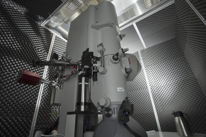
New specimen supports solve decades-long question on how to use liquid helium temperatures in cryo-EM to reduce radiation damage

Electron cryo-microscopy (cryo-EM) is an invaluable tool for mapping the structures of biological molecules. It works by rapidly freezing samples in a thin layer of ice using liquid ethane, loading them onto a mesh-like grid to provide a stable support surface, and scanning them with an electron beam. However, structure determination at atomic levels has always been limited by radiation damage.
Over the past four decades, there have been numerous attempts to reduce the effects of radiation damage by cooling specimens with liquid helium, which is significantly colder than liquid nitrogen. Though this does reduce radiation damage by restricting the diffusion of damaging free radicals, all attempts using liquid helium have not produced any improvement in single-particle structure determination. Chris Russo’s group, in the LMB’s Structural Studies Division, has identified the physical causes behind information loss in previous attempts with liquid helium and designed new equipment to overcome these. The results are structural determination with liquid helium cooling where every frame contains more information than that available using liquid nitrogen.
Samples cooled by liquid nitrogen are taken to a temperature of roughly 80 Kelvins (K), the equivalent of roughly -193 degrees Celsius. Liquid helium is substantially colder, meaning the specimens are at a temperature of 13 K (approximately -260 ˚C) inside the microscope. It is established that cooling crystals from target molecules at liquid helium temperatures halves the effects of radiation damage in comparison to liquid nitrogen, so it stands to reason that adapting cryo-EM to use liquid helium would increase the capturable information (known as the signal) and reduce unwanted variations or disturbances (known as noise). Joshua Dickerson, a PhD student and latterly a postdoc in Chris’s group, now a postdoc at the University of California, Berkeley, set out to understand the physics behind data quality loss in liquid helium cooled cryo-EM.
First, it was established that the principal cause for loss of information at liquid helium temperatures was movement of the specimen. If the specimen moves just a few Ångstroms during imaging, this causes blurriness and loss of spatial detail. At liquid nitrogen temperatures, using grids with small holes of 200 to 300 nanometres (nm) diameter is enough to eliminate movement caused by the electron beam, but at liquid helium temperatures image quality was still compromised.
To find the cause of this movement, the group examined the movement of gold nanoparticles in 300 nm diameter holed grids using identical imaging conditions at 81 K and 13 K. At 81 K there was no significant particle movement and the grid and the layer of frozen water covering it remained flat throughout. However, at 13 K the particles travelled away from the centre of the grid holes, and this rate of displacement increased in line with increased hole diameter. Additionally, once it reached 13 K, stress accumulated in the grid as the layer of frozen water contracted at differing rates, causing the grid to crinkle. As the specimen is exposed to the electron beam, the frozen water became an ultra-viscous fluid, allowing the grid to relax and resume its flattened shape.
Surprisingly, the group also established that when the 13 K specimen is irradiated by the electron beam, the structure of the frozen water fundamentally changes. Instead of contracting as was previously thought, it was found that the amorphous frozen water actually expands, and will eventually buckle in each small hole. This explains the increase in distance between particles noted at 13 K compared to 81 K.
To remedy these issues, the group decided that the most straightforward solution would be to decrease the diameter of the holes in the support grids. They designed new grids with 100 nm holes, and found that using them in tandem with a 300 nm diameter electron beam eliminated the movement of the specimen. The smaller holes prevented any movement of the frozen water, and the decreased beam diameter meant a minimal area around each hole was irradiated, which prevented movement of the grid.
To test the efficacy of this new equipment, the group determined six structures at 13 K, and replicated these studies with the original cryo-EM setup at 81 K. Every frame captured at 13 K showed reductions in radiation damage and exhibited an improved signal-to-noise ratio in comparison to the 81 K structures. Ultimately, this illustrates that more structural information can be gathered using liquid helium. This improvement opens the possibilities of using cryo-EM to solve structures of far smaller proteins and molecules.
Looking to the future, this work has also highlighted important considerations in further equipment development for cryo-EM at lower temperatures. Though the 100 nm holed grids are key, large macromolecules and proteins that have a propensity to aggregate at the edge of holes are not well suited to such small hole diameters. This, and the requirement for minimal irradiation of the water and the grid, are crucial points to review when developing new cryo-microscopes and specimen supports optimised for liquid helium temperatures.
This work was funded by UKRI MRC, Astex Pharmaceuticals and the Herchel Smith fund.
Further references
Reducing the effects of radiation damage in cryo-EM using liquid helium temperatures. Dickerson, J.L., Naydenova, K., Peet, M.J., Wilson, H., Nandy, B., McMullan, G., Morrison, R., Russo, C.J. PNAS
Chris’s group page
Ultracool microscopy yields a sharper look at proteins – Science News
Previous Insight on Research articles
Low energy cryo-EM to widen accessibility for fast and accurate structure determination
Scaling-up grid manufacture to solve bottleneck in cryo-EM
Freezing molecules completely still for cryo-EM