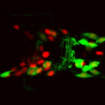
The connectome of an animal is the comprehensive map, or wiring diagram, of all the neural connections in the brain. However, an important challenge is how to make sense of this information. The nematode worm Caenorhabditis elegans, still the only animal for which the entire connectome has been described, illustrates the problem. Although it has only 302 neurons, these make thousands of connections. Even in such a simple animal, understanding the structure of the network, and the information flow within it is extremely challenging, so how can we hope to do this for connectomes that are many orders of magnitude larger? In this work Bill Schafer’s group in the LMB’s Neurobiology Division worked with Petra Vértes from the University of Cambridge Department of Psychiatry and Albert-Laszlo Barabasi‘s group from the Northeastern University Center for Complex Network Research in the US to apply control theory, normally used by engineers, to predict neural function and pathways of information flow in a real nervous system.
Petra Vértes and Albert-Laszlo Barabasi‘s group used principles of control theory to determine the structural controllability of the C. elegans body muscles (i.e. how many individual muscles could in principle be independently controlled) from an input signal from specific sensory neurons. They then determined which neurons in the network would reduce that controllability if they were eliminated from the network. To test the predictions, Denise Walker in Bill’s group used a laser microbeam to specifically kill individual neurons predicted to be important for controllability, as well as some related neurons not predicted to affect controllability as negative controls. Then Yee Lian Chew, also in Bill’s group, used an automated tracking microscope to record the movements of the operated worms, and used machine vision approaches to analyse their behaviour. Remarkably, the control theory predictions were confirmed, with ablations of neurons required for full controllability having specific effects on behaviour that were not seen in control animals.
This approach can be scaled up to probe much larger neural networks, so it should be possible to use it to probe the structure and function of bigger brains such as our own. Mapping the human connectome could lead to major advances in our understanding of what makes us human and provide the basis for future studies of abnormal brain circuits in many neurological and psychiatric disorders. In order to effectively modulate a human brain network to treat cognitive deficits (e.g. using deep brain stimulation to treat Parkinson’s disease), it is necessary to understand how signals from one part of the brain influence targets elsewhere, and what are the critical links that allow these signals to reach their targets. This is an area of intense research, and huge technical advances have been made in the collection and analysis of the imaging data needed to achieve this. This work provides new and powerful theoretical tools to allow us to interpret these brain maps and understand the how the nervous system works at the most fundamental, cellular level.
This work was funded by the MRC, the Wellcome Trust, EMBO, EU Horizon2020 and the John Templeton Foundation.
Further references:
Paper in Nature
Bill’s group page
Petra Vértes’ group page
Albert-Laszlo Barabasi‘s group page