
LMB Image Game
This is an exciting scientific image from the LMB. What do you think it looks like? What could it be showing?
Click on the image to find out, then click on the arrow to see the next image.
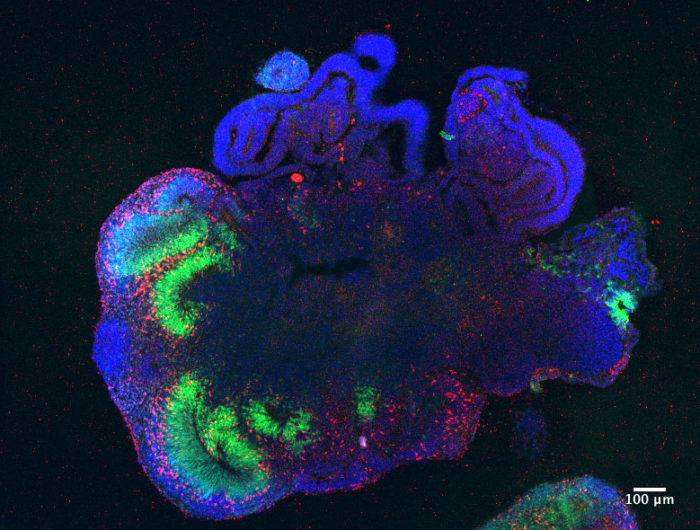
This is a ‘mini brain’ created by Madeline Lancaster’s lab in the LMB’s Cell Biology Division. Madeline’s lab uses stem cells from skin samples which are then bathed in nutrients and factors to trigger their development into neurons. The cells are then planted in a dense protein gel and amazingly as the cells replicate and grow, they organise themselves into a tiny version of our own brains – cerebral organoids, or ‘mini brains’. Madeline’s group is using these organoids to better understand brain development, including the changes in brain structure which are associated with conditions such as autism. 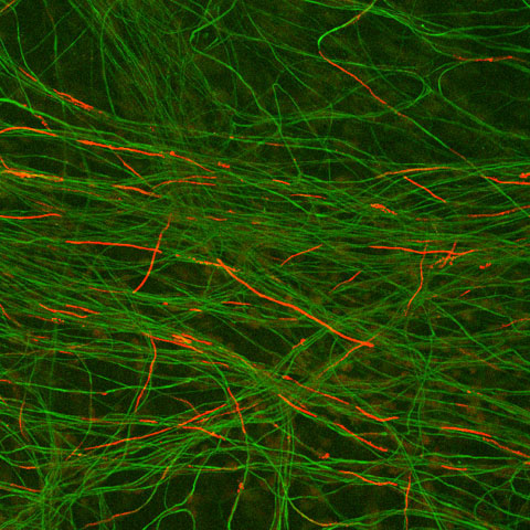
This is an image of some neurons, long cells which carry nervous signals around the body. The red areas show the presence of myelin which is an insulating sheath that surrounds nerves and increases the speed at which nervous signals travel. If myelin isn’t made properly, this can result in disease. The image was taken by scientists in Anne Bertolotti’s group in the LMB’s Neurobiology Division. The scientists are investigating drugs which can help when myelin isn’t made correctly, due to misfolding of myelin proteins, and may result in treatments for neurodegenerative diseases. 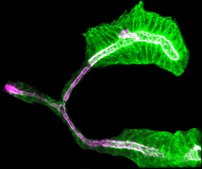
This is an image of a salivary gland from a fruit fly (Drosophila), taken in Katja Röper’s lab in the LMB’s Cell Biology Division. The gland consists of big secretory cells which make the saliva and small flat cells which line the duct carrying the saliva to the fly’s mouth. In the image, the cells are green and the duct is purple. Katja’s group studies the development of tubular organs such as the salivary glands from their initial existence as a flat sheet of cells to the complex tubular organ you see here. Knowledge gained from investigating tube development in the fruit fly can help inform our understanding of similar developmental processes in vertebrates, like ourselves. 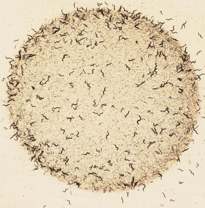
This is a photo, taken in Mario de Bono’s lab in the LMB’s Cell Biology Division, of lots of tiny 1mm long nematode worms (C. elegans) eating their lunch! Usually the worms cluster when they feed, but these worms have a mutation which means they behave differently. Mario’s group are using the worms to investigate the mechanisms underlying the behaviour. Scientists use ‘model organisms’, such as C. elegans, in their research because they are simple, easy to keep and help us understand biological processes which can be relevant to humans. 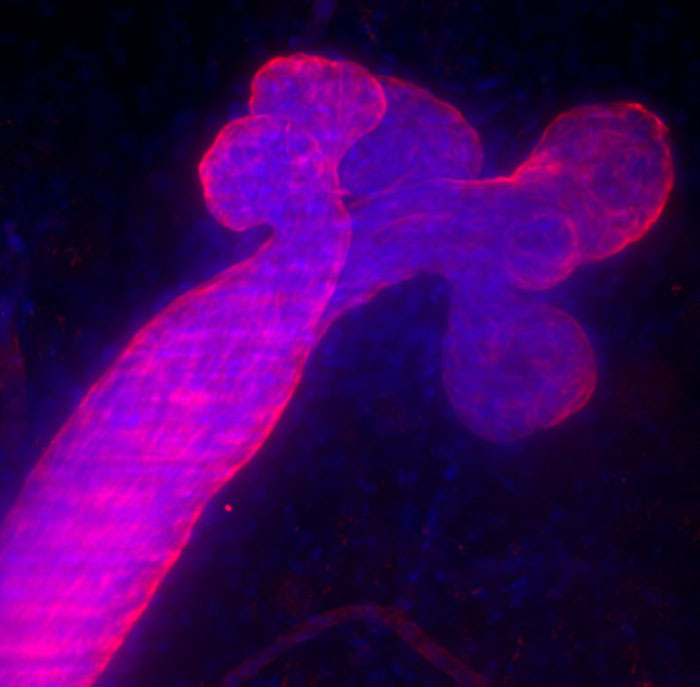
This is an image of mammary gland tissue, taken by Mathias Pasche in the LMB’s Cell Biology Division. Milk is made by the round structures (bulbs) in the top right of the image and then travels down the tube (milk duct) which points towards the bottom left corner of the image. This image was taken using special microscopy techniques which allow scientists to look deeper into the tissue sample, leading to improved awareness of how the tissue is organised into different regions. This work is helping scientists better understand the reasons for breast cancer and where different types of cancer have their origin.