Amphiphysin
Bringing your curves to the bar
Amphiphysin has an N-terminal lipid interaction, dimerisation and membrane bending BAR domain, a middle clathrin and adaptor binding domain (CLAP) and a C-terminal SH3 domain. There are 2 mammalian amphiphysins with similar overall structure, and a ubiquitous splice form of amphiphysin 2 that does not contain the CLAP domain. They are implicated in clathrin-mediated endocytosis by virtue of their binding to dynamin, synaptojanin clathrin and the AP2 adaptor complex. However they can also function in the muscle in the formation and organisation of the T-tubule network and likely in membrane bending events in other cell types.

Bananna-shapped BAR domain: dimerisation and membrane curvature-sensing domain. Crystal structrue from Drsoophila muscle amphiphysin
Drosophila Amphiphysin BAR domain (Peter et al. 2003)(pdf)
amphiphysin SH3 domain
Amphiphysin2 SH3 domain (Owen et al 1998)

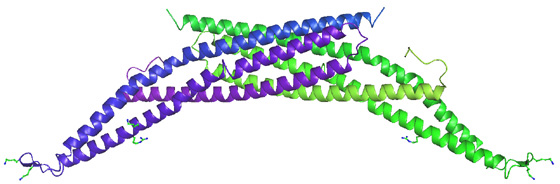
Amphiphysin was first implicated in the recruitment of dynamin by de Camilli and colleagues. Evidence later accumulated that amphiphysin may indeed be the recruitment protein for dynamin to sites of clathrin-mediated endocytosis. Despite the low affinity of the amphiphysin-SH3 domain for dynamin (Owen et al 1998 EMBO J.) the interaction is likely to be considerably stronger in vivo where amphiphysin is a dimer and dynamin is an oligomer (see Wigge et al 1997 MBC).

What makes amphiphysin a good candidate protein for dynamin recruitment? In brain extract amphiphysin is found in a 1:1 stoichiometry with dynamin and this interaction is inhibited by phosphorylation and both proteins are dephosphorylated on stimulation of exo-endocytosis. The amphiphysin SH3 domain when overexpressed in COS7 cells is a potent inhibitor of dynamin action when compared alongside other SH3 domains which binds to dynamin (Grb2, Spectrin and PLCg) (see Wigge et al 1997 Current Biol.). The SH3 domain also blocks endocytosis in the Lamprey synapse (Shupliakov et al 1997) leading to the accumulation of endocytic profiles with 'necks' along the plasma membrane. The potency of the isolated SH3 domain is likely due to its ability to disassembly dynamin multimers ( Owen et al 1989 EMBO J.). The binding site for amphiphysin has been carefully mapped to a sequence ProSerArgProAsnArg in the PRD of dynamin. Peptides that specifically inhibit the dynamin-amphiphysin interaction inhibit clathrin-mediated endocytosis. A mutation in a critical lipid binding residue of the PH domain (Lys535Ala) of dynamin leads to a dominant negative phenotype in cells (see Vallis et al 1999) but this can be partially rescued by a second mutation of the amphiphysin binding sequence in the PRD (see Vallis et al 1999) thus showing that the protein that interacts with this sequence plays an important role.
Apart from a dynamin, amphiphysin also independently binds to clathrin, AP2 adaptors and to lipids and dynamin co-localises with amphiphysin to clathrin positive puncta. This does not mean that this is the only dynamin recruitment protein for clathrin-mediated endocytosis because Drosophila amphiphysin is not expressed in nerve terminals and thus another protein (perhaps endophilin or DAP160) must perform this function. The Drosophila amphiphysin has no clathrin, and AP2 adaptor binding sites and does not bind to Drosophila dynamin. Deletion of the Drosophila amphiphysin gene results in a strong muscular phenotype (where it is highly expressed) rather than an endocytic phenotype. Amphiphysin in the muscle is implicated in making transverse t-tubule, consistent with its ability to tubulate membranes (Razzaq et al 2001 Genes and Development). This function of amphiphysin is also likely important in mammalian clathrin-mediated endocytosis where it may help to form the necks of endocytic vesicles.