EpsinR: an AP1/clathrin interacting protein

involved in vesicle trafficking

EpsinR is a ubiquitous homologue of epsin1. It is a 70kDa CCV enriched protein that binds to PtdIns(4)P, clathrin and AP1 adaptors (see paper). See also EpsinR home page

The images in these web pages are supplementary to those published in Mills et al., J. Cell Biol. 160: 213-222 (2003)
Myc-EpsinR with endogenous TGN46 and AP1 in COS cells

In the image to the left there are 2 cells transfected with Myc-epsinR and 3 cells not transfected. In the left cells endogenous AP1 is finely punctate and co-localises with the more tubular stain of endogenous TGN46. In the right transfected cell both the perinuclear AP1 stain and TGN46 stain are less compact and thus the co-localisation with epsinR is easier to visualise. We see that many of the perinuclear epsinR puncta co-stain for TGN46 and the AP1 complex. In non transfected cells overlapping TGN46 and AP1 are pink.

Click image to view individual channels or proceed to galleries of images using the links below.

A TGN localisation for a proportion of the epsinR is supported by the co-fractionation with TGN markers during organelle purification and the inhibitory effect of overexpression on trafficking of cathepsinD (see paper).

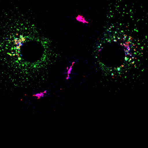
Click links below to view image galleries

Endogenous epsinR expression

Comparison of epsin1 and epsinR

Myc-epsinR (WT) overexpression and colocalisation with markers

Myc-epsinR D34G+R67L (lipid binding mutant)

Myc-epsinR L10E (proposed lipid conformation mutant)

Myc-epsinR D342R+D422R (clathrin binding mutant)

Myc-epsinR D34G+R67L + D342R+D422R (lipid and clathrin binding mutant)

EpsinR-GFP and ENTH domain overexpression

Sedimentation equilibrium analysis (.pdf)

EpsinR home page

McMahon lab home page