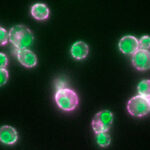
By combining fluorescence microscopy and electron tomography, Wanda Kukulski’s lab in Cell Biology Division has visualised protein structures that bridge contact sites between the endoplasmic reticulum and plasma membrane in yeast, in their native environment i.e. within the cell.
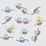
New insights into the architecture of organelle contact sites, and the sites’ roles in cellular lipid fluxes
New understanding of protein abundance control
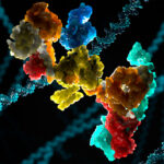
Decade-long collaboration results in the first structure of the Fanconi anaemia core complex
Our DNA contains all of the information required to tell a cell what it needs to do, but it is constantly being damaged. This damage can cause severe problems, making repair processes hugely important. One common type of DNA damage, known as crosslinking, involves links forming inappropriately between two nucleotide letters.
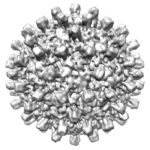
Fast, simple, accessible and affordable: The future of cryo-EM
Is it possible to improve imaging of purified biological specimens in electron cryo-microscopy (cryo-EM) while also reducing its cost? The latest proof-of-principle paper from Chris Russo’s group says yes, and indicates that the answer lies in reducing the electron energy in the cryo-EM from the current standard of 300 or 200 kiloelectron volts (keV) to 100 keV.
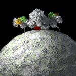
Structural insights into control of cell growth by nutrient availability
Control of cell division is crucially important, as unregulated cell division is a hallmark of cancer. mTORC1 protein kinase is an ancient enzyme complex and master regulator of growth and metabolism that integrates signals relating to nutrient availability, energy, and growth factors. Activation of mTORC1 is driven by proteins called Rags that sense nutrient abundance.
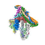
How chromosomes are bound to be separated in cell division
When our cells divide, it is important that the pairs of chromosomes are correctly segregated, as errors in this process cause serious problems. For over a century, kinetochores have been recognised as the critical cellular structures responsible for attaching the chromosomes to the microtubules that direct this chromosomal segregation. However, how exactly kinetochores recognise the centromere, the central point that links the two halves of a chromosome, has been a long-standing question.