Crystallisation and X-ray

A high quality crystal will amplify the reflections of an intense, monochromatic, beam of light. The resulting diffraction patterns are processed mathematically to solve the crystal structure and observe details of the corresponding molecules with atomic resolution. This is the fundamental principle of X-ray crystallography applied to structural biology for a better understanding of the functions of proteins, RNA, DNA and their complexes. Another application is drug discovery since compounds with high affinity will be observed in binding pockets.

However, the crystallisation of purified biological samples is associated with low success, notably because of the unstable and flexible nature of the macromolecules investigated. In addition, the right crystallisation conditions cannot be anticipated. In order to increase the yield of high quality crystals, multiple sample variants must be produced and analysed, guided by the support of other scientific facilities, such as biophysics and mass spectrometry.
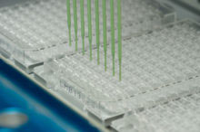
For the in-house base of 60-80 users to screen efficiently a large number of crystallisation conditions with a minimal amount of sample, automated protocols for robotic nanoliter experiments have been implemented and optimised. There are seven liquid-handling robots (some integrated into two fully automated systems) and 3 rooms dedicated to assessment and storage of crystallisation experiments. The main strategy is based on a large initial screen made of 2,112 conditions in pre-filled MRC plates.
There are two X-ray generators in the X-ray facility: a Rigaku FR-E+ SuperBright generator and a Rigaku Micromax-007HF generator, each equipped with two detectors. LMB researchers also have access to the Diamond synchrotron in Harwell and the European Synchrotron Radiation Facility in Grenoble (ESRF), including remote data collection, as well as other synchrotrons for specialised purposes.
In parallel with giving support, experts at the LMB also participate to progress methods in macromolecular X-ray crystallography by regularly creating and developing tools, such as the MORPHEUS protein crystallization screen, the MOSFLM data processing software, the REFMAC refinement program and the graphics-based model building and visualisation program COOT.
The crystallisation facility is run by Fabrice Gorrec and the X-ray facility is run by Dom Bellini.
Material to produce the figure on top was provided by Roger Williams’ group, PNAC (crystal structure of ESCRT-II complex).
Selected Papers
Gorrec, F. and Löwe, J. (2018)
Automated Protocols for Macromolecular Crystallization at the MRC Laboratory of Molecular Biology.
JoVE 131, 55790.
Gorrec, F. (2009)
The Morpheus protein crystallisation screen.
J Appl Cryst 7, 1035–1042.
Powell, H.R., Battye, T.G.G., Kontogiannis, L., Johnson, O. and Leslie, A.G.W. (2017)
Integrating macromolecular X-ray diffraction data with the graphical user interface iMosflm.
Nat Protocols 12, 1310–1325.
Murshudov, G.N., Skubák, P., Lebedev, A.A., Pannu, N.S., Steiner, R.A., Nicholls, R.A., Winn, M.D., Long, F. and Vagin, A.A.(2011)
REFMAC5 for the refinement of macromolecular crystal structures.
Acta D 67, 355–367.
Emsley, P., Lohkamp, B., Scott, W.G. and Cowtan K. (2010)
Features and development of Coot.
Acta D 66, 486–501.