While fully sequenced genomes are available for many important experimental organisms, a major challenge has been to identify the functions of the genes identified. A method for phenotyping that is both high-throughput, so all an organism’s genes can be phenotyped, and high-content, so inferences about gene function can be made with precision, has been required.
The nematode worm, C. elegans, is a major experimental model for neuroscience, as well as aging and development.
Identifying behavioural functions for genes has produced a key resource for neuroscience research
The smell of food ‘wakes up’ the zebrafish visual system
New research from Leon Lagnado’s group in the LMB’s Neurobiology Division shows how food-related smells ‘re-tune’ zebrafish vision by making the retina more sensitive to moving objects, such as the prey that zebrafish eat.
The way the brain processes information from one sense depends on the activity of other senses. For instance, we all know that to listen closely to some music, it often helps to shut one’s eyes.
Work on ubiquitination reveals insights into disease
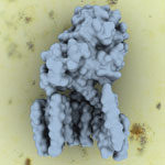
Recent work carried out by David Komander’s group in the LMB’s Protein and Nucleic Acid Chemistry Division, and published in two separate papers, has provided insight into human disease and the role played by ubiquitination, a process that affects many fundamental cellular processes.
Work in David’s group aims to understand the cellular machinery for specific ubiquitin (Ub) assembly, disassembly, and binding on a structural and biochemical level.
Insights into spliceosomal activation and molecular pathology of retinitis pigmentosa eye disease
Work published in the journal Structure by Kiyoshi Nagai’s group in the LMB’s Structural Studies Division, has provided further detailed information on the structure and role of proteins at the active site of the spliceosome, and may also help to explain the molecular pathology of the eye disease, retinitis pigmentosa type 13 (RP13).
Watching neurons transmit visual information
Work in Leon Lagnado’s group in the LMB’s Neurobiology Division is showing how synapses transmit visual signals in the retina of zebrafish. The group designed fluorescent proteins that light up when synapses are active and made transgenic zebrafish expressing these proteins in retinal neurons. They then used a multiphoton microscope to directly observe the activity of synapses in the retina of live fish as they responded to different visual stimuli.
New mechanism in the body’s timekeeping revealed
Our cycle of sleep and wakefulness is controlled by a daily (circadian) body clock in our brain. When this cycle happens in a regular way people function well, but when this cycle is disturbed it can lead to a severely disrupted life. The suprachiasmatic nucleus (SCN) is part of the body clock and individual neurons of the SCN contain their own 24-hour clock, but they operate best when connected together in their neural circuit and run in synchrony.