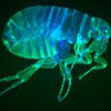
Mesolens microscopy (2009)
The LMB’s Brad Amos is a key developer of confocal microscopy at the LMB and the creator of the Mesolens – a unique microscope with a giant lens that can examine thousands of cells and the detail inside each cell at the same time.
The Mesolens will be able to capture detail in organisms that are too big to be examined satisfactorily by existing microscopes, offering a deeper insight into areas such as cancerous tissues and the cortex of the brain.
Developing faster, more detailed images

The Mesolens microscope was developed in response to the need to examine larger and larger tissue samples, such as the 6mm, early stage, genetically modified mouse embryos used by scientists to help identify future treatments for diseases such as cancer.
The confocal microscope, the current gold standard in biomedical imaging, is successful because it allows biologists to obtain an image from inside a thick specimen. However, these microscopes only allow small images to be obtained – in order to build an image of a 6mm object hundreds of these images would need to be pieced together.
With the Mesolens, scientists can obtain all the information in a single image. As well as being faster, this new type of imaging can reach deeper into solid tissues, such as embryos or tumours.
The Mesolens offers doctors the possibility of vastly improved diagnostic examinations. The technology has already opened up new possibilities: for macrophotography in biomedical optics, in imaging neurones and mammalian embryos, and in aiding bioluminescence studies. It is also likely to be of value in mineralogy, materials, food and forensic science.
In 2009 Brad Amos set up a private company, Mesolens Ltd, under the aegis of the MRC, to develop the technology. A test centre for Mesoscopy has been set up in the University of Strathclyde, and commercial production of microscopes based on this technology is planned.