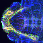
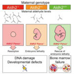
A cross divisional collaboration at the LMB between the groups of Simon Bullock, in Cell Biology, and Andrew Carter, in Structural Studies, has provided new insight into the activation of the large molecular motor dynein, a critical component of the transport system that operates within cells.
The cells within living organisms contain an elaborate transport system that moves different components to the right part of the cell at the right time.
How to get your (dynein) motor running
Cryo-EM reveals mammalian protein export machinery
A collaborative team from LMB’s Cell Biology and Structural Studies Divisions has visualized the mammalian protein synthesis and export machinery at near-atomic resolution. The new research helps explain how secreted proteins, such as hormones, can cross an otherwise impermeable membrane to exit the cell.
It has long been appreciated that cells communicate with each other via proteins that are either secreted or embedded in the cell’s surface.
How does biology make tubes?
Insights into how the Fanconi Anaemia core complex activates DNA repair
Research carried out by Eeson Rajendra from Lori Passmore’s group in the LMB’s Structural Studies Division, in close collaboration with KJ Patel from the LMB’s PNAC Division, has brought together LMB expertise in protein biochemistry and genetics to study the disease Fanconi Anaemia (FA). For the first time, they have isolated the intact FA core complex, and demonstrated which subunits are essential for monoubiquitination of FANCD2, which initiates the repair of damaged DNA in cells.
Novel lipid kinase structure lays the foundation for a new class of drugs
A collaboration between Roger Williams’ group here in the LMB’s Protein and Nucleic Acid Chemistry Division and Kevan Shokat’s group at the University of California, San Francisco has provided insight into potential targets for the design of a new class of anti-viral drugs.
Enteroviruses cause diseases including polio; hand, foot and mouth disease and the common cold, and there are currently no anti-viral treatments available to combat them.
Graphene: stronger than steel and now suitable for biological electron microscopy
Lori Passmore and Chris Russo from the LMB’s Structural Studies Division have discovered how to modify the new material graphene using low-energy hydrogen plasmas, to allow it to bind proteins. This discovery makes it suitable for use in electron microscopy to solve protein structures.
Graphene, which was only discovered in 2004, is a two-dimensional carbon sheet that is one of the strongest and thinnest materials known.