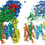
A research team from the LMB’s Cell Biology division, working with colleagues from the Structural Studies division, has revealed how cells are able to find and tag for degradation the partially synthesised proteins generated when ribosomes occasionally stall.
Cells make more than a hundred thousand new proteins every minute. Once in a while, one of the ribosomes making these proteins stalls, leaving an unfinished protein fragment.
Unfinished proteins: how to find a needle in the haystack
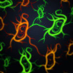
Insight into bacterial cell division: Architecture of the FtsZ ring
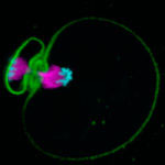
When a bacterial cell divides, the cell membrane and cell envelope have to pinch together in the middle of the cell to separate it into two daughter cells. A ring of proteins called the divisome constricts, cleaving the cell in two. The protein FtsZ is a crucial component of this ring and many FtsZ subunits join together in a chain forming long filaments. These FtsZ filaments are anchored to the membrane by another protein, FtsA, so that the membrane also constricts when the FtsZ ring closes.
Golden grids for electron cryo-microscopy
Recent exciting advances in electron cryo-microscopy (cryo-EM) have allowed scientists to find very detailed structures of some proteins. Still, determining the structure of many proteins remains too difficult for cryo-EM, as the images are too noisy to use for structure determination. Lori Passmore and Chris Russo from the LMB’s Structural Studies Division have designed new specimen support grids, made of pure gold, that improve the microscope image quality.
Signposts for organelle identity – new Rab GTPase effectors found
Cells contain specialised membrane-bound compartments called organelles, which are vital to the cell as they allow it to separate different biochemical reactions that otherwise might interfere with each other. To function correctly, these intracellular compartments need to recruit proteins from the cytoplasm, and since every organelle has a specific role, each one needs a particular set of proteins.
Structure of human dynein shows the powerstroke mechanism
Dyneins are a family of motor proteins that move along microtubules powered by chemical energy from ATP. Andrew Carter and his group in the LMB’s Structural Studies Division have solved the structure of a dynein protein bound to a chemical that mimics the shape of ATP, and have shown for the first time how the dynein can ‘walk’ along the microtubule.
Dynein proteins carry various important cargos to different parts of the cell, and are crucial to correct cell function.