
LMB Fellows
The LMB Fellows scheme aims to facilitate or enhance long-term collaborations between scientists from across the world and LMB and its groups. LMB Fellowships are sponsored by a group, they are time limited and provide the Fellows with access to the LMB and its facilities, making the Fellows highly valued members of the LMB’s scientific community.
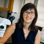


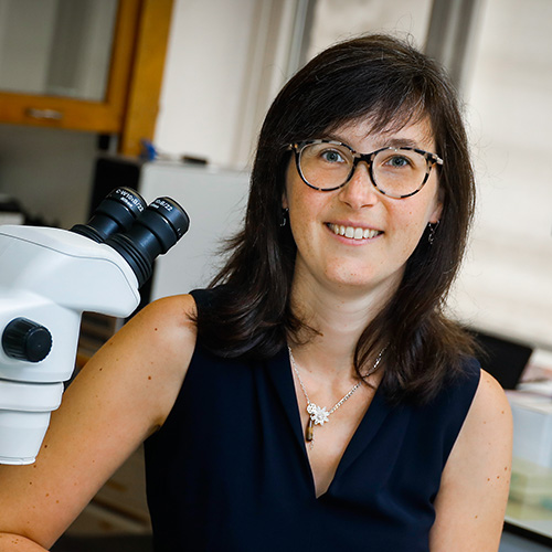
Isabel Beets
Sponsor:- Bill Schafer, Neurobiology
Isabel Beets is Associate Research Professor at the Biology Department of the University of Leuven, Belgium. Her group is interested in the diversity of neuromodulators, such as neuropeptides, that regulate behaviour and experience-based learning. Their aim is to understand the system-wide organisation of the neuropeptide signalling network and how it controls behavioural plasticity. Her team is investigating these questions in the well-defined nervous system of the nematode C. elegans.A core approach is to map neuropeptide-receptor interactions and the experience-dependent activity of these signalling networks using reverse pharmacology and genetically-encoded GPCR sensors. With Bill Schafer’s group they are working to determine the complete network of ‘wireless’ peptidergic communication – the neuropeptidergic connectome – of the C. elegans nervous system and to use this map to better understand how behaviour is modulated by experience.
- Beets, I., Zels, S., Vandewyer, E., Demeulemeester, J., Caers, J., Baytemur, E., Courtney, A., Golinelli, L., Hasakioğulları, İ., Schafer, W.R., Vértes, P.E., Mirabeau, O., Schoofs, L. (2023)
System-wide mapping of peptide-GPCR interactions in C. elegans.
Cell Rep 42(9): 113058 - Watteyne, J., Peymen, K., Van der Auwera, P., Borghgraef, C., Vandewyer, E., Van Damme, S., Rutten, I., Lammertyn, J., Jelier, R., Schoofs, L., Beets, I. (2020)
Neuromedin U signaling regulates retrieval of learned salt avoidance in a C. elegans gustatory circuit.
Nat Commun 11(1): 2076 - Beets, I., Janssen, T., Meelkop, E., Temmerman, L., Suetens, N., Rademakers, S., Jansen, G., Schoofs, L. (2012)
Vasopressin/oxytocin-related signaling regulates gustatory associative learning in C. elegans.
Science 338(6106): 543-5
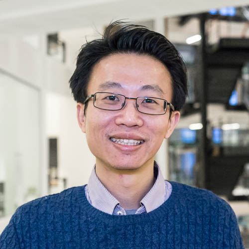
Patrick Cai
Sponsor:- Julian Sale, PNAC
Prof. “Patrick” Yizhi Cai received a bachelor degree in Computer Science in China, a master degree in Bioinformatics from University of Edinburgh in the UK, and a PhD in Genetics, Bioinformatics and Computational Biology from Virginia Tech in the USA. Prof. Cai had his postdoctoral fellowship under Jef Boeke in the Johns Hopkins University School of Medicine. Prof. Cai serves as a senior scientific consultant to Beijing Genomics Institute, and is the first Autodesk Distinguished Scholar. From 2013 to 2017, Prof. Cai had his own research group at the University of Edinburgh with a prestigious Chancellor’s Fellowship, and his lab focuses on Computer Assisted Design for Synthetic Biology, NeoChromosome design and synthesis in the yeast, and DNA assembly automation. In the summer of 2017, Prof. Cai moved to the University of Manchester as the new chair professor in synthetic genomics. Prof. Cai co-founded Edinburgh Genome Foundry, the International Centre for Synthetic Genomics at BGI, and the GP-Write China Centre at Chinese Academy of Sciences.
- Luo, Z., Yu, K., Xie, S., Monti, M., Schindler, D., Fang, Y., Zhao, S., Liang, Z., Jiang, S., Luan, M., Xiao, C., Cai, Y., Dai, J. (2021)
Compacting a synthetic yeast chromosome arm.
Genome Biol 22(1): 5 - Dai, J., Boeke, J.D., Luo, Z., Jiang, S., Cai, Y. (2020)
Sc3.0: revamping and minimizing the yeast genome.
Genome Biol 21(1): 205 - Liu, W., Luo, Z., Wang, Y., Pham, N.T., Tuck, L., Pérez-Pi, I., Liu, L., Shen, Y., French, C., Auer, M., Marles-Wright, J., Dai, J., Cai, Y. (2018)
Rapid pathway prototyping and engineering using in vitro and in vivo synthetic genome SCRaMbLE-in methods.
Nat Commun 9(1): 1936
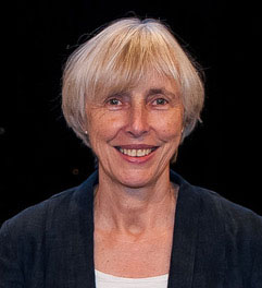
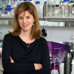
Caroline Dean
Sponsor:- Lori Passmore, Structural Studies
Caroline Dean is based at the John Innes Centre (Norwich). Over many years her lab has elucidated the mechanisms through which plants register seasonal temperature signals to time their development. This question has taken her into the study of conserved co-transcriptional and epigenetic mechanisms underpinning gene silencing. Genetic screens had revealed that regulation of the major Arabidopsis floral repressor gene (FLC) linked 3’ processing/transcription termination to changes in the local chromatin environment. These include chromatin modifications that determine levels of transcriptional output, promote cold-induced epigenetic switching, and feed back to influence RNA 3’processing/ transcription termination. Recently, a collaboration with the Passmore lab has shown that physical association of the CPSF phosphatase module with chromatin modifiers is a core part of this transcription-coupled silencing mechanism. Their collaboration will now actively investigate the interconnections between transcription termination with the delivery of a changed chromatin environment, linking structural insight with in vivo significance.
- Fang, X., Wu, Z., Raitskin, O., Webb, K., Voigt, P., Lu, T., Howard, M., Dean, C. (2020)
The 3' processing of antisense RNAs physically links to chromatin-based transcriptional control.
Proc Natl Acad Sci U S A 117(26): 15316-15321 - Fang, X., Wang, L., Ishikawa, R., Li, Y., Fiedler, M., Liu, F., Calder, G., Rowan, B., Weigel, D., Li, P., Dean, C. (2019)
Arabidopsis FLL2 promotes liquid-liquid phase separation of polyadenylation complexes.
Nature 569(7755): 265-269
- Mateo-Bonmatí E., Fang X., Maple R, Fiedler M., Passmore L.A., and Dean C. (2023)
The CPSF phosphatase module links transcription termination to chromatin silencing
BioRxiv
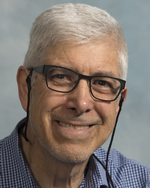
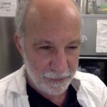
Paul Edelstein
Sponsor:- Lalita Ramakrishnan, Cell Biology
Paul Edelstein is a clinical microbiologist and infectious diseases consultant who is a foremost expert in Legionnaires’ disease, including its clinical, microbiological and pathogenesis aspects. He is also expert in clinical microbiology diagnostics, antimicrobial resistance and antibiotics, having served as the Director of the Clinical Microbiology Laboratory of the Hospital of the University of Pennsylvania for three decades. For the last decade, he has been involved in research on tuberculosis pathogenesis and epidemiology. Paul is now an emeritus professor of Pathology and Laboratory Medicine at the Perelman School of Medicine of the University of Pennsylvania, where he continues to teach clinical microbiology fellows. He is also an Honorary Senior Research Fellow in the Department of Medicine of the University of Cambridge. He is a frequent visitor to the LMB, collaborating closely with Lalita Ramakrishnan’s lab and more recently has also begun to advise Tanmay Bharat’s research.
- Fan, J., Hale, V.L., Lelieveld, L.T., Whitworth, L.J., Busch-Nentwich, E.M., Troll, M., Edelstein, P.H., Cox, T.M., Roca, F.J., Aerts, J.M.F.G., Ramakrishnan, L. (2023)
Gaucher disease protects against tuberculosis.
Proc Natl Acad Sci U S A 120(7): e2217673120 - Lake, M.A., Adams, K.N., Nie, F., Fowler, E., Verma, A.K., Dei, S., Teodori, E., Sherman, D.R., Edelstein, P.H., Spring, D.R., Troll, M., Ramakrishnan, L. (2023)
The human proton pump inhibitors inhibit rifampicin efflux and macrophage-induced rifampicin tolerance.
Proc Natl Acad Sci U S A 120(7): e2215512120 - Whitworth, L.J., Troll, R., Pagán, A.J., Roca, F.J., Edelstein, P.H., Troll, M., Tobin, D.M., Phu, N.H., Bang, N.D., Thwaites, G.E., Thuong, N.T.T., Sewell, R.F., Ramakrishnan, L. (2021)
Elevated cerebrospinal fluid cytokine levels in tuberculous meningitis predict survival in response to dexamethasone.
Proc Natl Acad Sci U S A 118(10)

Tom Ellis
Sponsor:- Jason Chin, PNAC
Tom Ellis is Professor of Synthetic Genome Engineering in the Bioengineering Department of Imperial College London and an Associate Faculty member of the Wellcome Trust Sanger Institute. Tom obtained his PhD in Cambridge studying the molecular sequence recognition at promoter regions. He then established a career in synthetic biology, leading a group since 2010 that develops tools and technologies for designing and assembling diverse genetic programs into microbial cells including yeasts. Since 2013, he has been part of the Synthetic Yeast Genome (Sc2.0) Project, an international collaboration rewriting the S. cerevisiae genome from computationally-designed synthetic DNA fragments. In collaboration with the Chin and Sale labs in PNAC, Prof Ellis is now developing the methods to achieve genome assembly from synthetic DNA in more complex cells, with a particular focus on chromosome synthesis in human cell lines.
- Ostrov, N., Beal, J., Ellis, T., Gordon, D.B., Karas, B.J., Lee, H.H., Lenaghan, S.C., Schloss, J.A., Stracquadanio, G., Trefzer, A., Bader, J.S., Church, G.M., Coelho, C.M., Efcavitch, J.W., Güell, M., Mitchell, L.A., Nielsen, A.A.K., Peck, B., Smith, A.C., Stewart, C.N., Tekotte, H. (2019)
Technological challenges and milestones for writing genomes.
Science 366(6463): 310-312 - Shaw, W.M., Yamauchi, H., Mead, J., Gowers, G.F., Bell, D.J., Öling, D., Larsson, N., Wigglesworth, M., Ladds, G., Ellis, T. (2019)
Engineering a Model Cell for Rational Tuning of GPCR Signaling.
Cell 177(3): 782-796.e27 - Blount, B.A., Gowers, G.F., Ho, J.C.H., Ledesma-Amaro, R., Jovicevic, D., McKiernan, R.M., Xie, Z.X., Li, B.Z., Yuan, Y.J., Ellis, T. (2018)
Rapid host strain improvement by in vivo rearrangement of a synthetic yeast chromosome.
Nat Commun 9(1): 1932
Gillian Griffiths
Sponsor:- Emmanuel Derivery, Cell Biology
Gillian Griffiths is a Wellcome Trust Principal Research Fellow based at the Cambridge Institute for Medical Research. Her research is focused on understanding the cell biology of cytotoxic T lymphocytes, key cells of the immune system that destroy virally infected and cancer cells. She identified a novel role for the centrosome in directing secretion of modified secretory lysosomes and has identified many of the proteins involved in this process by studying immunodeficiencies. Recent research has taken advantage on new advances in high resolution imaging to study the remarkable polarisation that these cells undergo when they encounter their targets. She is currently collaborating with Manu Derivery to use micropatterned surfaces to follow T cell polarisation and James Manton in using Light Sheet microscopy.
- Gawden-Bone, C.M., Frazer, G.L., Richard, A.C., Ma, C.Y., Strege, K., Griffiths, G.M. (2018)
PIP5 Kinases Regulate Membrane Phosphoinositide and Actin Composition for Targeted Granule Secretion by Cytotoxic Lymphocytes.
Immunity 49(3): 427-437.e4 - Richard, A.C., Lun, A.T.L., Lau, W.W.Y., Göttgens, B., Marioni, J.C., Griffiths, G.M. (2018)
T cell cytolytic capacity is independent of initial stimulation strength.
Nat Immunol 19(8): 849-858
Wanda Kukulski
Sponsor:- Madeline Lancaster, Cell Biology
Wanda Kukulski is Professor of Biochemistry at the Institute of Biochemistry and Molecular Medicine at the University of Bern, Switzerland. Her group is interested in the diversity of specialised membrane architectures that drive cellular functions. At present, their aim is to understand the molecular organisation and function of organelle contact sites, and dynamic reorganisations of mitochondrial membranes. The core approach is to visualise membrane shape and topology as well as protein assemblies associated with cellular membranes by correlative microscopy and electron cryo-tomography. With Madeline Lancaster’s group they are working on combining correlative microscopy with cerebral organoid technology. They aim to visualise molecular architectures in neuronal tissue, which will allow specific questions on organelle membranes in neurons to be addressed.
- Hoffmann, P.C., Bharat, T.A.M., Wozny, M.R., Boulanger, J., Miller, E.A., Kukulski, W. (2019)
Tricalbins Contribute to Cellular Lipid Flux and Form Curved ER-PM Contacts that Are Bridged by Rod-Shaped Structures.
Dev Cell 51(4): 488-502.e8 - Ader, N.R., Hoffmann, P.C., Ganeva, I., Borgeaud, A.C., Wang, C., Youle, R.J., Kukulski, W. (2019)
Molecular and topological reorganizations in mitochondrial architecture interplay during Bax-mediated steps of apoptosis.
Elife 8 - Bharat, T.A.M., Hoffmann, P.C., Kukulski, W. (2018)
Correlative Microscopy of Vitreous Sections Provides Insights into BAR-Domain Organization In Situ.
Structure 26(6): 879-886.e3

Paul Lehner
Sponsor:- Felix Randow, PNAC
Paul Lehner is a Wellcome Trust Principal Research Fellow and Honorary Consultant in Infectious Diseases at the CITIID Institute in the Jeffrey Cheah Biomedical Centre, Cambridge. His research focuses on the use of genetic and quantitative proteomic approaches to understand how viruses interact with and manipulate their host cells. His application of forward-genetic screens discovered the Human Silencing Hub ‘HUSH’, the critical transcriptional epigenetic repressor complex for silencing both newly integrated retroviruses and mobile endogenous retrotransposons. His collaboration with Felix Randow will take advantage of their complementary expertise to gain mechanistic insights into microbial pathogenesis using genetic and proteomic discovery platforms. They anticipate the discovery of common themes by which different pathogens exploit cellular pathways to overcome cell autonomous immunity and evade cellular recognition.
- Tchasovnikarova, I.A., Timms, R.T., Douse, C.H., Roberts, R.C., Dougan, G., Kingston, R.E., Modis, Y., Lehner, P.J. (2017)
Hyperactivation of HUSH complex function by Charcot-Marie-Tooth disease mutation in MORC2.
Nat Genet 49(7): 1035-1044 - Greenwood, E.J., Matheson, N.J., Wals, K., van den Boomen, D.J., Antrobus, R., Williamson, J.C., Lehner, P.J. (2016)
Temporal proteomic analysis of HIV infection reveals remodelling of the host phosphoproteome by lentiviral Vif variants.
Elife 5 - Tchasovnikarova, I.A., Timms, R.T., Matheson, N.J., Wals, K., Antrobus, R., Göttgens, B., Dougan, G., Dawson, M.A., Lehner, P.J. (2015)
GENE SILENCING. Epigenetic silencing by the HUSH complex mediates position-effect variegation in human cells.
Science 348(6242): 1481-1485

Ben Luisi
Sponsor:- Sjors Scheres, Structural Studies.
Ben Luisi is a structural biologist based in the Department of Biochemistry, Cambridge. He and his research group study RNA metabolism related to the posttranscriptional control of gene expression. He is working with Tom Dendooven on rules for ribonucleoprotein mediated regulation. In collaboration with Michal Wandel and Felix Randow, his group explored therapeutic oligonucleotides against SARS-CoV-2 during the first lockdown. They also study regulation and assembly of efflux pump assembles and have collaborated with Sjors Scheres and his colleagues to elucidate the structure of a pump by cryoEM.
- Dendooven, T., Sinha, D., Roeselová, A., Cameron, T.A., De Lay, N.R., Luisi, B.F., Bandyra, K.J. (2021)
A cooperative PNPase-Hfq-RNA carrier complex facilitates bacterial riboregulation.
Mol Cell 81(14): 2901-2913.e5 - Lulla, V., Wandel, M.P., Bandyra, K.J., Ulferts, R., Wu, M., Dendooven, T., Yang, X., Doyle, N., Oerum, S., Beale, R., O'Rourke, S.M., Randow, F., Maier, H.J., Scott, W., Ding, Y., Firth, A.E., Bloznelyte, K., Luisi, B.F. (2021)
Targeting the Conserved Stem Loop 2 Motif in the SARS-CoV-2 Genome.
J Virol 95(14): e0066321 - Fitzpatrick, A.W.P., Llabrés, S., Neuberger, A., Blaza, J.N., Bai, X.C., Okada, U., Murakami, S., van Veen, H.W., Zachariae, U., Scheres, S.H.W., Luisi, B.F., Du, D. (2017)
Structure of the MacAB-TolC ABC-type tripartite multidrug efflux pump.
Nat Microbiol 2: 17070

Liz Miller
Sponsor- Ramanujan Hegde, Cell Biology
My group collaborates with Manu Hegde’s group on our shared interests in secretory and membrane protein biogenesis. The long-standing question of my lab has been to reveal how export of secretory and membrane proteins from their site of synthesis in the endoplasmic reticulum (ER) is influenced by protein folding. We make use of the Hegde lab’s in vitro translation systems to reconstitute protein biogenesis, observe direct interactions with protein chaperones and export factors, and measure protein secretion. By understanding the molecular details of these protein-protein interactions, we hope to develop small molecules that selectively inhibit secretion of some proteins but leave the rest of the secretome intact.
- Maldutyte, J., Li, X.H., Gomez-Navarro, N., Robertson, E.G., Miller, E.A. (2025)
ER export via SURF4 uses diverse mechanisms of both client and coat engagement.
J Cell Biol 224(1) - Gomez-Navarro, N., Maldutyte, J., Poljak, K., Peak-Chew, S.Y., Orme, J., Bisnett, B.J., Lamb, C.H., Boyce, M., Gianni, D., Miller, E.A. (2022)
Selective inhibition of protein secretion by abrogating receptor-coat interactions during ER export.
Proc Natl Acad Sci U S A 119(31): e2202080119 - Stancheva, V.G., Li, X.H., Hutchings, J., Gomez-Navarro, N., Santhanam, B., Babu, M.M., Zanetti, G., Miller, E.A. (2020)
Combinatorial multivalent interactions drive cooperative assembly of the COPII coat.
J Cell Biol 219(11) - Gomez-Navarro, N., Melero, A., Li, X.H., Boulanger, J., Kukulski, W., Miller, E.A. (2020)
Cargo crowding contributes to sorting stringency in COPII vesicles.
J Cell Biol 219(7)
Kim Nasmyth
Sponsor- Jan Löwe, Structural Studies
Kim Nasmyth currently holds the Whitley Chair at the Department of Biochemistry, University of Oxford and is a fellow of Trinity college. He was a PhD student in Mitchison’s lab in Edinburgh (1974-77), a post doc in Seattle Washington (1978-1980), a Robertson research fellow at Cold Spring Harbor (1980-81), and a member of staff at the MRC laboratory for molecular biology in Cambridge (1982-87) before moving to the Research Institute of Molecular Pathology (I.M.P) in Vienna, where he was a senior scientist from 1988 to 1997 and director from 1997 to 2006. His lab identified the cohesin complex that regulates the topology of eukaryotic chromosomes during interphase and holds sister chromatids together following their genesis during S phase. They also elucidated the mechanism by which cohesion is destroyed at the metaphase to anaphase transition, namely cleavage of cohesin’s kleisin subunit by separase, a thiol protease activated through ubiquitinylation of its inhibitory chaperone securin by the Anaphase-promoting complex or cyclosome. Kim’s lab has collaborated closely with that of Jan Löwe for nearly twenty years. Their analyses of cohesin’s structure have led to the notion that it holds sister DNAs together by entrapping them inside a tripartite ring formed by pairwise interactions between its Smc1, Smc3, and kleisin subunits. Their most recent work together has revealed aspects of the complex that enable it to compact DNAs through loop extrusion, in particular the ATP-dependent clamping of DNAs onto top of the Smc1/3 ATPase domains by a regulatory subunit called Scc2 (Nipbl). Kim’s work has been recognized by several awards, including the Gairdner foundation prize and the Breakthrough prize. He is a fellow of the Royal Society, a member of the Austrian Academy of Sciences, and a foreign honorary member of the American Academy of Arts and Sciences.
- Maldutyte, J., Li, X.H., Gomez-Navarro, N., Robertson, E.G., Miller, E.A. (2025)
ER export via SURF4 uses diverse mechanisms of both client and coat engagement.
J Cell Biol 224(1) - Gomez-Navarro, N., Maldutyte, J., Poljak, K., Peak-Chew, S.Y., Orme, J., Bisnett, B.J., Lamb, C.H., Boyce, M., Gianni, D., Miller, E.A. (2022)
Selective inhibition of protein secretion by abrogating receptor-coat interactions during ER export.
Proc Natl Acad Sci U S A 119(31): e2202080119 - Stancheva, V.G., Li, X.H., Hutchings, J., Gomez-Navarro, N., Santhanam, B., Babu, M.M., Zanetti, G., Miller, E.A. (2020)
Combinatorial multivalent interactions drive cooperative assembly of the COPII coat.
J Cell Biol 219(11) - Gomez-Navarro, N., Melero, A., Li, X.H., Boulanger, J., Kukulski, W., Miller, E.A. (2020)
Cargo crowding contributes to sorting stringency in COPII vesicles.
J Cell Biol 219(7)
David Owen
Sponsor- Sean Munro, Cell Biology
David Owen is a Wellcome Trust Principal Research Fellow based at the Cambridge Institute for Medical Research. In joint efforts with a number of other laboratories, his group use an integrated structure/in vitro/in vivo approach to study membrane trafficking along the endocytic system of mammalian cells. This involves trying to understand the molecular recognition processes that occur within, and allow the formation of, the protein coats of tubular vesicular carriers, which move between the cell surface, endosomes and degradative endolysosomes. In collaboration with John Briggs’ group, the structures of minimal AP2/clathrin and retromer coats have been determined by cryo electron tomography: both systems are now being expanded to include more coat components. In collaboration with David Neuhaus’ NMR group, the regulation of both processes is being investigated using a combination of NMR, X-ray crystallography and high-resolution live cell imaging.
- Wrobel, A.G., Kadlecova, Z., Kamenicky, J., Yang, J.C., Herrmann, T., Kelly, B.T., McCoy, A.J., Evans, P.R., Martin, S., Müller, S., Salomon, S., Sroubek, F., Neuhaus, D., Höning, S., Owen, D.J. (2019)
Temporal Ordering in Endocytic Clathrin-Coated Vesicle Formation via AP2 Phosphorylation.
Dev Cell 50(4): 494-508.e11 - Kovtun, O., Leneva, N., Bykov, Y.S., Ariotti, N., Teasdale, R.D., Schaffer, M., Engel, B.D., Owen, D.J., Briggs, J.A.G., Collins, B.M. (2018)
Structure of the membrane-assembled retromer coat determined by cryo-electron tomography.
Nature 561(7724): 561-564
Ewa Paluch
Sponsor- Kate McDole, Cell Biology
Ewa Paluch currently holds the Chair of Anatomy in the Department of Physiology, Development and Neuroscience at the University of Cambridge. Her lab combines molecular and cell biology, biophysics, quantitative imaging and modelling to investigate the principles underlying cellular morphogenesis. The lab’s research directions span cell surface mechanics regulation, the control of cell shape during cell division and migration, and the cross-talk between cell mechanics, cell shape and cell fate. Research questions Ewa plans to explore in collaboration with LMB scientists are how the structural and mechanical properties of actin networks constrain possible actin organisation states, how these in turn define the landscape of shapes displayed by cells, and how cells navigate their morphogenetic landscape during cellular state transitions.
- Truong Quang, B.A., Peters, R., Cassani, D.A.D., Chugh, P., Clark, A.G., Agnew, M., Charras, G., Paluch, E.K. (2021)
Extent of myosin penetration within the actin cortex regulates cell surface mechanics.
Nat Commun 12(1): 6511 - De Belly, H., Stubb, A., Yanagida, A., Labouesse, C., Jones, P.H., Paluch, E.K., Chalut, K.J. (2021)
Membrane Tension Gates ERK-Mediated Regulation of Pluripotent Cell Fate.
Cell Stem Cell 28(2): 273-284.e6 - Chugh, P., Clark, A.G., Smith, M.B., Cassani, D.A.D., Dierkes, K., Ragab, A., Roux, P.P., Charras, G., Salbreux, G., Paluch, E.K. (2017)
Actin cortex architecture regulates cell surface tension.
Nat Cell Biol 19(6): 689-697
Gerald Rubin
Sponsor:- Greg Jefferis, Neurobiology
Gerald Rubin is a Senior Group Leader at the Howard Hughes Medical Institute’s Janelia Research Campus. His research focuses on developing an experimental approach to neurobiology based on the comprehensive identification and manipulation of individual cell types and circuit components. His laboratory uses the tools and methods they develop to study the neuronal circuits in Drosophila underlying learning and memory, sleep regulation, visual perception, sensory integration, and aggression. With Greg Jefferis’ group they are working, as part of a large international collaboration, to determine the complete wiring diagram – or connectome – of the adult Drosophila nervous system and to use that wiring diagram to gain insights into how the brain controls behavior.
- Schretter, C.E., Aso, Y., Robie, A.A., Dreher, M., Dolan, M.J., Chen, N., Ito, M., Yang, T., Parekh, R., Branson, K.M., Rubin, G.M. (2020)
Cell types and neuronal circuitry underlying female aggression in .
Elife 9 - Wu, M., Nern, A., Williamson, W.R., Morimoto, M.M., Reiser, M.B., Card, G.M., Rubin, G.M. (2016)
Visual projection neurons in the lobula link feature detection to distinct behavioral programs.
Elife 5 - Aso, Y., Rubin, G.M. (2016)
Dopaminergic neurons write and update memories with cell-type-specific rules.
Elife 5
Jeanne Salje
Sponsor:- Buzz Baum, Cell Biology
Jeanne Salje is a Wellcome Trust Senior Research Fellow at the Cambridge Institute for Medical Research, an Assistant Professor at the Department of Biochemistry and the Department of Pathology at Cambridge University, and an Honorary Visiting Research Fellow at the Mahidol Oxford Tropical Medicine Research Unit in Bangkok, Thailand. Her research focuses on understanding the fundamental biology of a group of bacteria that exclusively replicate inside living eukaryotic cells. These obligate intracellular bacteria cause a range of human and animal diseases, and the Salje lab is particularly focused on one called Orientia tsutsugamusi that causes the severe but neglected human disease scrub typhus. Research areas include mechanisms of bacterial growth and division, interactions with host cells, and disease pathogenesis.Dr. Salje is working with Buzz Baum to explore how an understanding of obligate intracellular bacteria informs our understanding of endosymbiosis and the modern eukaryotic cell.
- Atwal, S., Wongsantichon, J., Giengkam, S., Saharat, K., Pittayasathornthun, Y.J., Chuenklin, S., Wang, L.C., Chung, T., Huh, H., Lee, S.H., Sobota, R.M., Salje, J. (2022)
The obligate intracellular bacterium Orientia tsutsugamushi differentiates into a developmentally distinct extracellular state.
Nat Commun 13(1): 3603 - Atwal, S., Chuenklin, S., Bonder, E.M., Flores, J., Gillespie, J.J., Driscoll, T.P., Salje, J. (2021)
Discovery of a Diverse Set of Bacteria That Build Their Cell Walls without the Canonical Peptidoglycan Polymerase aPBP.
mBio 12(4): e0134221 - Mika-Gospodorz, B., Giengkam, S., Westermann, A.J., Wongsantichon, J., Kion-Crosby, W., Chuenklin, S., Wang, L.C., Sunyakumthorn, P., Sobota, R.M., Subbian, S., Vogel, J., Barquist, L., Salje, J. (2020)
Dual RNA-seq of Orientia tsutsugamushi informs on host-pathogen interactions for this neglected intracellular human pathogen.
Nat Commun 11(1): 3363
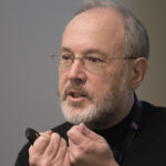
John Walker
Sponsor:- Michel Goedert, Neurobiology
Since 2013, John Walker has been Director Emeritus at the MRC Mitochondrial Biology Unit in Cambridge. In 1994, his work, started in 1981 in the LMB’s PNAC Division and continued throughout the intervening period, on characterizing the ATP synthase in the mitochondria (and also in chloroplasts and bacteria), led to the realisation that the energy released by the oxidation of dietary sugars and fats is coupled by a mechanical rotary mechanism to generate the 60 kg of ATP that each of us makes daily to sustain our lives. This work led to the award of the Nobel Prize in Chemistry in 1997. In the 1990s, he also defined the protein composition of another huge molecular assembly in mitochondria, known as complex I, and the localisation of the 44 constituent proteins within the complex. Complex I is a key component in the respiratory chain of enzymes that converts energy from food into a transmembrane proton motive force (pmf). This work provided essential foundations for more recent investigations by colleagues into its structure and function.
Since leaving the LMB in 1998, he has continued to delve deeper into the mechanism of the ATP synthase, and especially into the question of how rotation is generated from the pmf. His recent description in 2021 (with T. Spikes and M. Montgomery) of the structure of the complete dimeric mitochondrial ATP synthase provides crucial evidence that rotation is generated by the direct application of the pmf (predominantly a voltage) to the membrane domain of the enzyme’s rotor via a Grotthus water chain. John uses his knowledge on energy conversion for medical benefit, in 2021 establishing the structure of the mycobacterial ATP synthase as a target for developing new drugs against tuberculosis.
John is a Fellow of the Royal Society, and in 2012, he was awarded its Copley Medal. He is also a Fellow of the Academy of Medical Sciences, a Fellow of Sidney Sussex College, Cambridge, an Honorary Fellow of St Catherine’s College, Oxford, a Foreign Member of L’Accademia Nazionale dei Lincei, of the Royal Netherlands Academy of Arts and Sciences, and of The Royal Society of New Zealand, and a Foreign Associate of the US National Academy of Sciences.

Malcolm Weir
Sponsor:- Chris Tate, Structural Studies
Malcolm Weir is a biochemist and entrepreneur, currently working as an Independent Consultant in the field of drug discovery and early development. He started his career in Glaxo where he established structure-based drug design and led a broad range of discovery technologies. He then moved as CEO to the structural chemo/bioinformatics company Inpharmatica. In 2007, along with Chris Tate, Richard Henderson and Fiona Marshall he co-Founded Heptares as CEO and remained with it as Head of R&D after its sale to Sosei, leaving in 2022. He is strongly interested in the molecular biosciences and their application to find new medicines, and to this end is keen to work with scientists and investors to help start new biotechnology companies. He is a non-Exec Director of the drug discovery company Kesmalea Therapeutics Ltd.
He is Visiting Professor of Biochemistry at Imperial College London, has Hon DSc degrees from ICL and the University of Hertfordshire, is an Hon Member of the British Biophysical Society, a Fellow of the Royal Society of Chemistry and was a co-recipient of the 2015 Royal Society of Chemistry Malcolm Campbell Award for Biological Chemistry.
- Brown, A.J.H., Bradley, S.J., Marshall, F.H., Brown, G.A., Bennett, K.A., Brown, J., Cansfield, J.E., Cross, D.M., de Graaf, C., Hudson, B.D., Dwomoh, L., Dias, J.M., Errey, J.C., Hurrell, E., Liptrot, J., Mattedi, G., Molloy, C., Nathan, P.J., Okrasa, K., Osborne, G., Patel, J.C., Pickworth, M., Robertson, N., Shahabi, S., Bundgaard, C., Phillips, K., Broad, L.M., Goonawardena, A.V., Morairty, S.R., Browning, M., Perini, F., Dawson, G.R., Deakin, J.F.W., Smith, R.T., Sexton, P.M., Warneck, J., Vinson, M., Tasker, T., Tehan, B.G., Teobald, B., Christopoulos, A., Langmead, C.J., Jazayeri, A., Cooke, R.M., Rucktooa, P., Congreve, M.S., Weir, M., Tobin, A.B. (2021)
From structure to clinic: Design of a muscarinic M1 receptor agonist with potential to treatment of Alzheimer's disease.
Cell 184(24): 5886-5901.e22 - Jazayeri, A., Rappas, M., Brown, A.J.H., Kean, J., Errey, J.C., Robertson, N.J., Fiez-Vandal, C., Andrews, S.P., Congreve, M., Bortolato, A., Mason, J.S., Baig, A.H., Teobald, I., Doré, A.S., Weir, M., Cooke, R.M., Marshall, F.H. (2017)
Crystal structure of the GLP-1 receptor bound to a peptide agonist.
Nature 546(7657): 254-258 - Jazayeri, A., Doré, A.S., Lamb, D., Krishnamurthy, H., Southall, S.M., Baig, A.H., Bortolato, A., Koglin, M., Robertson, N.J., Errey, J.C., Andrews, S.P., Teobald, I., Brown, A.J., Cooke, R.M., Weir, M., Marshall, F.H. (2016)
Extra-helical binding site of a glucagon receptor antagonist.
Nature 533(7602): 274-7