• Skip to primary navigation
  • Skip to main content
  • Skip to primary sidebar
MRC Laboratory of Molecular Biology

MRC Laboratory of Molecular Biology

One of the world's leading research institutes, our scientists are working to advance understanding of biological processes at the molecular level - providing the knowledge needed to solve key problems in human health.

  • Home
  • About LMB
  • Research
  • Research Groups
  • Students
  • Recruitment
  • Life at the LMB
  • Achievements
  • News & Events
Home > Research Leaders > H to M > Kate McDole
Kate McDole cb

Kate McDole

Morphogenesis of the early mammalian embryo

kmcdole@mrc-lmb.cam.ac.uk

Development begins with a single cell of limitless potential giving rise to all of the different cell types, tissues, and organ systems that comprise an adult animal. How cells organize to build tissues and how those tissues are sculpted to form organs is largely unknown. While there have been significant advances in the organoid and stem cell fields in differentiating specific cell types and populations, we are not yet able to replicate the physical environment required to build more elaborate structures.  In the McDole lab, we use the mouse embryo to study how mechanical forces shape complex three-dimensional structures out of simple cell populations. We use a combination of cutting-edge live-imaging, biochemical techniques, computational methods, genetics and biophysics to dissect the role of forces during development.

The ability to watch development as it unfolds reveals how tissues form, however mammalian development has been notoriously difficult to visualize. Embryos are highly sensitive to environmental and culture conditions when removed from the mother, and in addition are extremely photosensitive.

We have developed an advanced light-sheet microscope to gently and comprehensively image mouse embryo development at single-cell resolution over a course of days. With this system and a suite of computational tools, we follow changes in cell fate, visualize the organization of tissue structures, and measure the forces involved in shaping those structures. Starting with the endoderm, we are examining how and where forces are generated to turn a relatively simple, single cell layered structure into the primordial gut tube, as well as how this process is coordinated with the development of neighbouring germ layers such as the heart tube and neural ectoderm.

One of the ultimate aims of the lab is to generate a comprehensive force-map of development in the early embryo that, when coupled with our existing knowledge of gene expression and cell fate maps, will allow us to generate computational models that can be used to design and engineer better 3D culture or synthetic tissue culture systems, enabling us to grow more complex structures in a dish and one day build working tissues and organs.


Video Player
https://www2.mrc-lmb.cam.ac.uk/wordpress/wp-content/uploads/MouseDevs.mp4
00:00
00:00
00:00
Use Up/Down Arrow keys to increase or decrease volume.

Two-day long time-lapse imaging of an H2BeGFP expressing mouse embryo from E6.5 d.p.c. to E8.5 d.p.c. (Top panel: distal view, bottom left panel: AP view, bottom right panel: lateral view).

Microscope image
Illustration of the Mouse SiMView microscope.
Embryo Diagram
Cartoon illustration of mouse development from E6.5 to E8.5, showing major tissue and organ structures such as the developing heart (purple), endoderm/foregut (green) and anterior epiblast/neural ectoderm (light blue).

Video Player
https://www2.mrc-lmb.cam.ac.uk/wordpress/wp-content/uploads/HeartForegutDevs.mp4
00:00
00:00
00:00
Use Up/Down Arrow keys to increase or decrease volume.

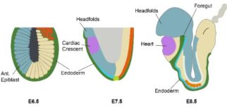
Time-lapse imaging of early cardiac development using an early
cardiac marker (Nkx2.5, in green) and a ubiquitous label (membrane
marker, in magenta). The cardiac crescent begins to come together
over the invaginating foregut pocket before converging at the midline
and forming the early heart tube.

Selected Papers

  • McDole K., Guignard L., Amat F., Berger A., Malandain G., Royer LA., Turaga SC., Branson K. & Keller PJ. (2018)
    In toto imaging and reconstruction of post-implantation mouse development at the single-cell level.
    Cell 175: 859-876.
  • McDole, K. and Zheng, Y. (2012)
    Generation and live imaging of an endogenous Cdx2 reporter mouse line.
    Genesis 50: 775-782.
  • McDole, K., Xiong, Y., Iglesias, P.A. and Zheng, Y. (2011)
    Lineage mapping the pre- implantation mouse embryo by two-photon microscopy, new insights into the segregation of cell fates.
    Dev. Biol. 355: 239-249.

Group Members

  • Sonia Agüera Gonzalez
  • Katharine Goodwin
  • Jenny Kretzschmar
  • Ewa Paluch
  • Henry Westmacott

Primary Sidebar

Research Leaders

  • A to G
    • Matteo Allegretti
    • Radu Aricescu
    • Diana Arseni
    • David Barford
    • Buzz Baum
    • Anne Bertolotti
    • Tanmay Bharat
    • Simon Bullock
    • Albert Cardona
    • Andrew Carter
    • Jason Chin
    • Emmanuel Derivery
    • Juliette Fedry
    • Michel Goedert
    • Joe Greener
    • Ingo Greger
  • H to M
    • Michael Hastings
    • Ramanujan Hegde
    • Philipp Holliger
    • Leo James
    • Gregory Jefferis
    • Joergen Kornfeld
    • Patrycja Kozik
    • Madeline Lancaster
    • Roni Levin Konigsberg
    • Jan Löwe
    • Kate McDole
    • Andrew McKenzie
    • Harvey McMahon
    • Liz Miller
    • Sean Munro
    • Garib Murshudov
  • N to S
    • Kelly Nguyen
    • John O’Neill
    • Lori Passmore
    • Lalita Ramakrishnan
    • Venki Ramakrishnan
    • Felix Randow
    • Jing Ren
    • Wes Robertson
    • Noe Rodriguez
    • Christopher Russo
    • Benjamin Ryskeldi-Falcon
    • Julian Sale
    • William Schafer
    • Sjors Scheres
    • Marta Shahbazi
    • John Sutherland
  • T to Z
    • Chris Tate
    • Marco Tripodi
    • Ana Tufegdžić Vidaković
    • Roger Williams
    • Joseph Yeeles
    • Suyang Zhang
    • Marta Zlatic
    • Jerome Zürcher
  • Emeritus
    • Brad Amos
    • Mariann Bienz
    • Tony Crowther
    • Phil Evans
    • Alan Fersht
    • Michael Gait
    • Richard Henderson
    • Rob Kay
    • John Kendrick-Jones
    • John Kilmartin
    • Peter Lawrence
    • Andrew Leslie
    • David Neuhaus
    • Hugh Pelham
    • Daniela Rhodes
    • Murray Stewart
    • Andrew Travers
    • Nigel Unwin
    • Greg Winter
  • LMB Fellows
  • Molecular Immunity Unit

Search

  • Privacy & Cookies
  • Contact Directory
  • Freedom of Information
  • Site Map
Find Us
©2025 MRC Laboratory of Molecular Biology,
Francis Crick Avenue, Cambridge Biomedical Campus, Cambridge CB2 0QH, UK. 01223 267000

The MRC is part of UK Research and Innovation

Contact Us

This site uses cookies. The LMB may use cookies to analyse how you use our website. We use external analysis systems which may set additional cookies to perform their analysis. These cookies (and any others in use) are detailed in our Privacy and Cookies Policy and are integral to our website. You can delete or disable these cookies in your web browser if you wish, but then our site may not work as it is designed. Ok