

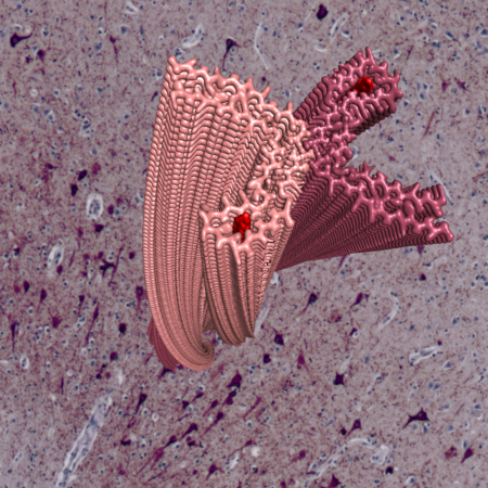
Ordered assembly of a small number of proteins within neurons and, in some cases, glia is a feature of neurodegenerative diseases. These proteins form amyloid structures enriched in repetitively stacked beta-strands. Mutations in the genes encoding these proteins lead to assembly and inherited neurodegenerative disease, demonstrating a causal role. Assemblies arise in discrete brain regions, from where they appear to propagate (spread and amplify) within connected brain networks, ultimately leading to neurodegeneration.
We work to understand the molecular basis of protein assembly into amyloid in neurodegenerative disease. Key questions that interest us are: How do assemblies form and propagate? How does this contribute to nerve cell degeneration? Why are only some cell populations in the brain vulnerable? Why does selective vulnerability differ between diseases?

To answer these questions, we combine structural and cell biology, using high-resolution electron microscopy with patient samples and advanced models of assembly in neuronal and glial cells. Electron cryo-microscopy (cryo-EM) of filament structures from human brains revealed that tau assemblies exist as disease-specific conformers, formed from conserved secondary structure motifs with different turn conformations and non-proteinaceous components. We are now studying how these structures relate to selective cell vulnerability by focusing on the molecular interactions between assemblies and brain cells.
Selected Papers
- Stephan Tetter, Diana Arseni, Alexey G. Murzin, Yazead Buhidma, Sew Y. Peak-Chew, Holly J. Garringer, Kathy L. Newell, Ruben Vidal, Liana G. Apostolova, Tammaryn Lashley, Bernardino Ghetti & Benjamin Ryskeldi-Falcon (2023)
TAF15 amyloid filaments in frontotemporal lobar degeneration
Nature, doi.org/10.1038/s41586-023-06801-2. - Scheres S, Ryskeldi-Falcon B, Goedert M. (2023)
Molecular pathology of neurodegenerative diseases by cryo-EM of amyloids.
Nature 621: https://doi.org/10.1038/s41586-023-06437-2 - Arseni D, Chen R, Murzin A G, Peak-Chew S Y, Garringer H J, Newell K L, Kametani F, Robinson A C, Vidal R, Ghetti B, Hasegawa M and Ryskeldi-Falcon B. (2023)
TDP-43 forms amyloid filaments with a distinct fold in type A FTLD-TDP
Nature, doi.org/10.1038/s41586-023-06405-w - Yang Y, Arseni D , Zhang W, Huang M , Lövestam S, Schweighauser M, Kotecha A, Murzin A G, Peak-Chew S Y, Macdonald J, Lavenir I, Garringer H J, Gelpi E, Newell K L, Kovacs G G, Vidal R, Ghetti B, Ryskeldi-Falcon B, Scheres S H W, and Goedert M (2022)
Cryo-EM structures of amyloid-β 42 filaments from human brains
Science, pp. 167-172 - Arseni D, Hasegawa M, Murzin AG, Kametani F, Arai M, Yoshida M, Ryskeldi-Falcon B. (2021)
Structure of pathological TDP-43 filaments from ALS with FTLD
Nature, doi: 10.1038/s41586-021-04199-3.
Group Members
- Diana Arseni
- Tiana Behr
- Heidy Chen
- Stephan Tetter
- Nikhil Varghese