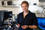

The McDole lab seeks to understand how complex 3D structures in the embryo are sculpted from initially homogenous cell populations. For example, how does an intricate, functional organ such as a beating heart arise from a uniform ball of cells? How do tubes involute and tissues fold, and what are the physical forces that drive these shape changes? Furthermore, how do cells themselves cope with different and changing mechanical environments, and how does this affect their gene expression and ultimate fate?
Many of these processes have never before been visualized or studied in a mamammalian embryo, and much of our understanding of the cell behaviors that drive early organogenesis remain a black box. Mammalian embryos are large, sensitive, and require complex culture requirements to sustain normal development outside of the uterus. In the McDole lab, we utilize advanced, adaptive light-sheet microscopy and a wide-range of specialized tools and methods to visualize development real-time. Coupled with advanced computational methods, we seek to quantiatively reconstruct early mammalian development and unveil the processes that shape tissues and organs, and uncover exciting new biology along the way.
Opportunities for PhD projects include 1) Using advanced light-sheet microscopy to peer into unseen areas of development 2) Investigating the mechanisms underlying the coordinated organization of the anterior side of the embryo – including the heart, brain, and early gut.
References
Cell 175(3): 859-876.e33 (2018)